Eliminate Risk – Expand Care
How newly established MRI safety protocols and training can dramatically improve care
Clinical Affairs | CU School of Medicine Mar 19, 2020
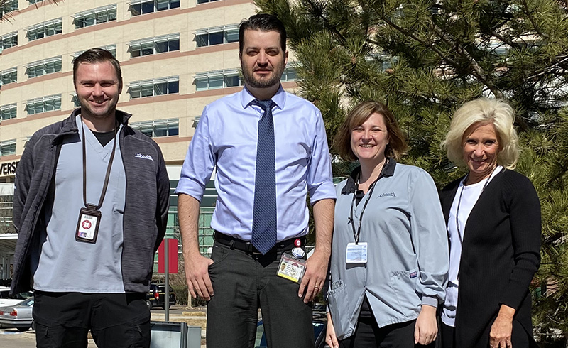
l-r, Paul Price, Justin Honce, Amber Seeman, Nancy Pritchard
A man is shot, and doctors at University of Colorado Hospital need to do an MRI to determine the best course of treatment. The only problem? The bullet is still lodged in his body, making an MRI potentially unsafe. Thanks to the work of Justin Honce, MD, Assistant Professor, Radiology-Neuroradiology, and his colleagues, getting the MRI is now possible.
Erring Too Far on the Side of Caution
An MRI, or magnetic resonance imaging scan, uses electromagnetic forces to create images of the inside the body. These forces do not harm human tissue but have the potential to affect foreign materials in the body, like pacemakers, defibrillators or metal screws and rods. Or bullets.
For a long time, this theoretical risk meant that people with such materials in their bodies could not have MRIs. Given the risk, the standard protocol was simply to say no.
But was it possible to evaluate the risk instead of simply saying no? This is the question Dr. Honce and other radiologists around the country were asking.
Opening Options without Compromising Safety
In 2015, Dr. Honce was looking broadly at all aspects of MRI safety at University Hospital and had the opportunity to receive training and certification in assessing the risks of MRIs when foreign materials are present. He knew he wanted to do this at University of Colorado Hospital.
He and his team began working with electro-physiology to develop a protocol*. “Having a systematic framework for evaluation of implanted devices and foreign bodies was the first step and the key,” said Dr. Honce. “That was the only way to maintain safety for patients while opening up MRI as a viable option.”
“Justin’s knowledge of MR and his dedication to MR safety combine to provide our patients with safe, timely, and outstanding quality care. UCH is fortunate to have Justin leading our MR endeavors,” says James Borgstede, MD, Vice Chair of Quality, Safety and Clinical Operations – Department of Radiology.
The risk assessment involves three lines of inquiry, based on the three different electromagnetic forces at work in an MRI:
- Magnetic force. The MRI is a big magnet with a large magnetic field. Will the magnet cause any metal inside the body to move?
- Radiofrequency energy. The MRI generates radiofrequency waves – this is the “resonance” part of magnetic resonance imaging. This energy creates heat in the body. Will the energy heat up a wire in the body and damage the tissue around it? If so, is there a way to redirect the wire or avoid it?
- Gradient or time-varying magnetic fields. These extra magnetic fields change the main magnetic field to allow for better resolution. They cause alternating electric currents to move back and forth, resulting in electrical stimulation. Will it cause electrical stimulation of the heart or brain?
If all three are cleared, it is safe for the patient to receive an MRI.
This protocol was rolled out in July 2018, and the hospital currently scans about 30 patients a month who would otherwise have gone without a needed MRI. University Hospital is currently one of the only institutions in Colorado that does this.
Putting the Right Team in Place
By its nature, an MRI is a multidisciplinary procedure. To ensure MRI safety, the hospital needed three key roles: the MR Medical Director, the MR Safety Expert and the MR Safety Officer. All three roles are board certified through the American Board of Magnetic Resonance Safety.
Dr. Honce is the current Medical Director, and his role is to consult with other physicians and answer technical questions. The MR Safety Expert is a medical physicist who advises on the device and the electromagnetic forces it uses, and tests different implants and foreign bodies to determine how the effect the MRI will have on them.
The MR Safety Officer is an MRI technologist; the hospital currently has eight trained Safety Officers led by MR Safety Manager Nancy Pritchard. They help other technologists to screen these higher-risk patients.
Training for Adoption of the New Practice
As with any new practice in medicine, the challenge was disseminating the information and training radiologic technologists to adopt the new process.
“It’s easy to say ‘no’ to patients with a risk. It’s harder to work up the case and put the effort forward, but that’s why training for technologists was so important,” explains Dr. Honce. “We needed radiologists and technologists to work together on this. Not all physicians are comfortable making decisions about MRI safety. The technologists can give radiologists the right information for decision making and offer their own recommendation as well.”
Training of technologists emphasized peer leadership and opportunities to practice working up cases under the guidance of a peer expert.
“This initiative exemplifies some of the most important factors for a successful new safety initiative,” said Read Pierce, MD, former Associate Director of Institute for Healthcare Quality, Safety, and Efficiency (IHQSE). “A carefully created protocol with comprehensive training and peer leadership – these increase the speed of adoption so we can give patients the best we have without delay.”
*Protocol based on a framework originally proposed by Emmanuel Kanal, MD, a neuroradiologist in Pittsburgh, PA.