Lab Services
Anatomic Imaging and Micromechanics
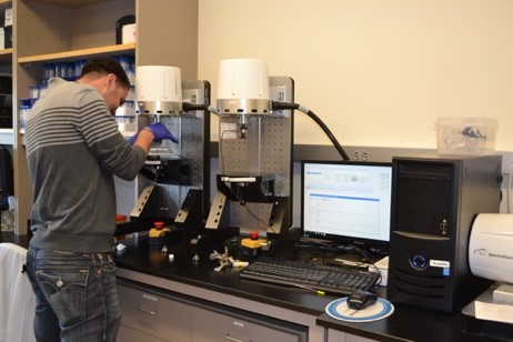
Microcomputed Tomography: Researchers in the Department of Orthopedics have access to a new, state-of-the-art high-resolution CT scanner (Scanco μCT50, installed March of 2018), equipped with a 12-stage robotic carousel. Image data and computational results are stored via NFS archival on institutional servers, guaranteeing no loss of data and rapid retrieval. Images are acquired and analyzed on an HP Integrity server (rx2800 i4) with a 13.84 (8 x 1.73) GHz CPU, 64 GB RAM, and 20 TB of local RAID storage. Researchers have installed customized routines for high-throughput batch scanning and automated selection of regions of interest.
Mechanical Testing Instruments: The Department of Orthopedics has installed new TA Instruments ELF 3200 series instrumentation, comprising 4 axial motors and 1 torsional motor under the control of two independent computer control systems. The electromagnetic actuators and contemporary software controls provide resolution in force (0.01 N) and displacement (1 μm) beyond that typically utilized in the mechanical testing of rodent bones.
Biomechanics Lab
The Biomechanics Lab is a state-of-the-art lab that encompasses biomechanical testing, device fabrication and evaluation, surgical simulation, and computer modeling. The lab performs research in all areas of orthopedic biomechanics.
 Tissue and Molecular Imaging
Investigators also have access to new equipment for processing and embedding tissue samples, with the ability to section tissue that is frozen, paraffin-embedded, or plastic-embedded. Key resources include:
- Sakura Tissue-Tek VIP6 AI Vacuum Infiltration Processor
- Sakura Tissue-Tek Tec6 Embedding Console System
- Leica CM1860 UV Cryostat with CryoJane Tape-Transfer System
- Leica Histo-Core AUTOCUT- Fully Motorized Microtome
- Leica Histo-Core MULTICUT- Semi-Motorized Microtome
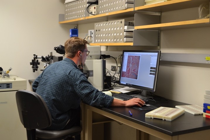
There are also resources and support for imaging of tissues or cell preparations using digital bright field or fluorescence and quantitative and histomorphometric image analysis. Current major equipment includes:
- Nikon Eclipse 80i upright fluorescence microscope with NIS Element AR software for standard fluorescence and light
microscopy - Olympus BX53 light and fluorescence microscope with an attached 20.7 Megapixel digital color camera and OsteoMeasureXP histomorphometry software V1.0.2.1 (OsteoMetrics, Inc, purchased in 2019) optimized for Windows 10 Pro workstations.
Skeletal Metabolism
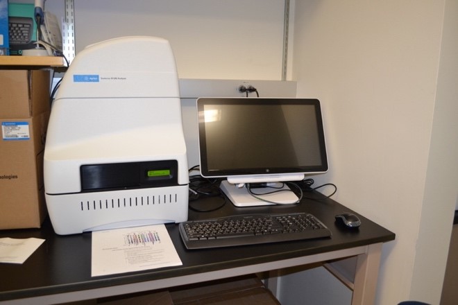
With the recent purchase of a Seahorse XFe96 Analyzer (Agilent), our research team is now able to measure the oxygen consumption rate and extracellular acidification rate of live cells, allowing the study of key cellular functions such as mitochondrial respiration and glycolysis.