Discussion and Pictures of Diabetic Retinopathy Lesions
| ||||||||||||||||||||||||||||||||
 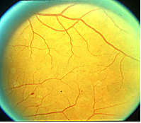 | Microaneurysms (Ma’s) – Ma’s are the first lesions appearing in diabetic retinopathy. Ma’s are physical dilations of the smallest intra-retinal blood vessels called capillaries. These lesions appear as small circular, red dots having distinct margins and are no larger than a blood vessel width at the disk margin. Good examples are seen in the following standard photograph 1: |
Figure 1. ETDRS Standard Photograph 1 showing Ma’s: Concentrate on the smallest red spots in the middle of the photo. | |
 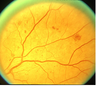 | Retinal Hemorrhages (H’s) – These lesions represent actual bleeding within the retina, and either are a result of ruptured Ma’s or when the above-mentioned capillaries become leaky enough to let blood out of the blood vessels (just as when we get a cut on our skin). These hemorrhages can be a variety of shapes including dot, blot and flame-shaped. They are usually larger than Ma’s (equal to or larger than a blood vessel at the disk margin), with uneven/indistinct edges and coloring. See the below standard photograph 2a: and concentrate on the larger red spots within the photograph. |
Figure 2. ETDRS Standard photograph 2a showing H/Ma’s: Concentrate on the larger red spots within the photograph. You may see some Ma’s in this photograph as well. This is because Ma’s and H’s commonly occur together (H/Ma’s). | |
 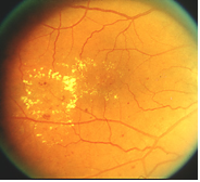 | Hard Exudates (HE’s) – These are white/yellow cholesterol deposits that usually originate from leaking Ma’s. HE’s are irregularly shaped, vary in size, are hard edged and often have a "fatty" appearance. HE can, and often are associated with fluid accumulation (retinal edema) within the retina. * HE and associated edema can occur adjacent to the macula, the part of the retina responsible for detailed, central vision. When the edema spreads into or very close the macula (clinically significant macular edema (CSME)), central vision can be affected and will cause the central vision to become constantly blurry. CSME can occur at any stage of retinopathy (non-proliferative or proliferative stages), and is usually treated with laser. |
Figure 3. ETDRS Standard photograph 4 showing HE’s in macula. | |
 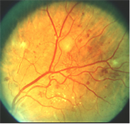 | Soft Exudates (SE) – aka "Cotton Wool Spots" - these lesions appear as white, feathery, fluffy or "cottony" spots. SE’s physically represent infarcts, or closures of capillaries, within the retina; however the physical locations of these lesions are in the very exterior layer of the retina (in the nerve fiber layer). Cotton Wool Spots often occur in association with IRMA (discussed below). |
Figure 4. ETDRS standard photograph 5 showing SE: Concentrate on the white areas just above the very center of the photo, at 3 o’clock (extreme right side of photo) and at 9:30 (just above the extreme left side of the photo). | |
 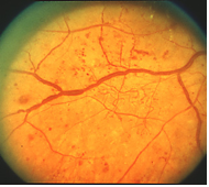 | Intraretinal Microvascular Anomalies (IRMA) – Occurring in the mid to late stages of non-proliferative retinopathy, these appear as spidery abnormal vessels that appear within the retina. They are typically contorted in appearance with sharp corners, often crossing over themselves and they normally do not cross over major veins or arteries. It is thought that IRMA physically represents either dilation of pre-existing capillaries or actual growth of new blood vessels within the retina. Venous Beading (VB) – Also occurring in the late stages of non-proliferative disease, VB occurs when the walls of major retinal veins loose their normal parallel alignment and begin to appear more like a string of sausages. Physically, IRMA represents a situation in which vein walls loose their elasticity and localized areas begin to dilate. This lesion is one of the strongest predictors of progression to proliferative disease. The above standard photograph represents this phenomenon very well. |
Figure 5. ETDRS standard photograph 6B Showing IRMA and VB: Proliferative Lesions | |
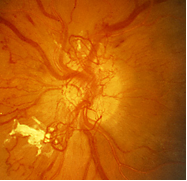
| New vessels (on the disc, NVD – or elsewhere, NVE) – aka "neovascularization"– NV are the first of the proliferative lesions to appear. The thought is that the retina is experiencing areas of hypoxia, or is "under-nourished". In an effort to compensate for this problem, the eye grows new vessels in the areas lacking circulation. The new vessels are very delicate and grow on top of the retina rather than in it, making them prone to leaking and hemorrhaging. They can cross over one or many major retinal blood vessels, but tend not to cross over themselves. They will often appear in a blossom like a flower bud with the outside part of the NV more dilated than the inner NV. They can also appear as stringy and drawn out, but this is less common. |
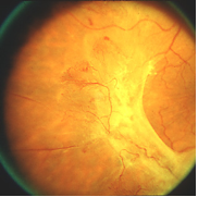
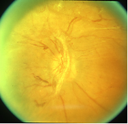
| Fibrous proliferation (on the disc, FPD - or elsewhere, FPE) - After the appearance of NV, the eye wants to protect these new vessels and give them support and structure to grow on. A white fibrous tissue grows either adjacent to or intertwined with the new vessels. This FP is very "sticky" and tends to adhere to the retina and NV. These adhesions can accidentally pull the NV and cause them to hemorrhage or can pull on the retinal and cause it to detach. |
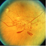
 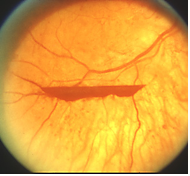 | Preretinal and vitreous hemorrhage (PRH, VH) – Occurs when the delicate NV leaks blood or is broken by the traction of fibrous proliferations. These two lesions are separated in name by their anatomical location. If this blood leaks into the potential space between the retinal and the internal limiting membrane, which lines the surface of the retina, it is called a preretinal hemorrhage. If the blood leaks into the vitreous or the posterior chamber, it is called a vitreous hemorrhage. |
Figure 10. ETDRS standard photograph 13 demonstrating a preretinal hemorrhage. | |
Acknowledgments:
| |

