Inner Visions
A Glimpse of What's Going on Inside
By Mark Couch
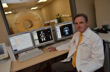
 (November 2013) A new research center at the School of Medicine will help scientists and physicians see some of the body’s inner workings, a crucial step toward better understanding and treatment of conditions that stubbornly resist sharing their secrets.
With updated body-scanning equipment and state-of-the-art laboratory techniques, the Colorado Translational Research Imaging Center (CTRIC) will capture images that yield some of those mysteries
“There was a very strong and diverse interest in expanding our research imaging capabilities, particularly in PET imaging,” Dodd says. “However, we had next to no existing PET researchers on campus. It was a chicken or the egg problem: No facility meant no researchers. We had to build the facility before we could create the program.”
And there was a lot that had to happen: purchasing a scanner that would be used only for research purposes, building a space to house it, establishing a laboratory where radiopharmaceuticals could be made, hiring the talent to run that lab, working to gain the federal government approvals necessary to use those specially created radiopharmaceuticals and establishing the protocols to prioritize the research requests
.The PET scan is particularly useful because it provides a peek at the body’s inner workings.
“It is an inside-out imaging technique, rather than outside-in,” Dodd says. “CT uses X-rays that originate outside the body and are passed through the body to create an image. With PET, you inject a
The person typically is injected with a radiopharmaceutical compound that includes a radioactive element, which shows up in the PET scan image. Glucose is a common base compound because it is fuel for cells. Active cells consume glucose and the radioactive element paired with the glucose emits rays that are measured by the PET scanner
“It’s always nice to build something from the ground up,” Smith-Jones says. “It’s a chance to leave a legacy.”
Smith-Jones works on the second piece of equipment, the cyclotron. It is located in a concrete bunker on the first floor of the Anschutz Inpatient Pavilion, behind doors with radiation warning signs. He uses the cyclotron to produce essential parts of the compounds used in research tests.
“We change one element into another,” Smith-Jones says. “It’s kind of like alchemy, like changing lead into gold, only we’re changing nitrogen into carbon.”
Though he quickly notes it’s not magic,
For example, by converting the element nitrogen-14 into carbon-11 in the cyclotron, Smith-Jones creates a radioisotope that can replace natural carbon-12 in
 “Most pharmaceutical compounds are intentionally made in amounts that will have an impact on biological function and treatment of the targeted disease,” Smith-Jones says. “In contrast, our compounds are made in very small amounts so that they don’t have any effect on the body, but can still be imaged by the PET scanner.”
For Huntington Potter’s study, for example, a specially designed
As the only imaging research center of its kind in the Rocky Mountain region, Dodd says scientists and physicians at the School of Medicine will now have the ability to conduct their research closer to home. And the school can nurture relationships with other institutions; researchers from CU-Boulder are conducting one of the pilot projects at CTRIC.
The price tag for such investments is significant: For CTRIC, the investment totals about $6 million.
Dodd said the center’s broad support led to funding from 14 medical school departments, centers
The funding does more than purchase equipment. It establishes a center with staff to make radiopharmaceuticals and manage the process of en-suring compliance with federal regulations. It creates a learning hub that sponsors a lecture series on advances in imaging. And it provides the startup funding for pilot projects that seek to investigate new research questions and generate pilot data for future grant funding.
“CTRIC provides significant new research opportunities for translational researchers on our campus from basic science through clinical trials,” Dodd says. “It provides a new view of the inner workings of health and disease and will lead to earlier and better treatments for our patients.”