Cellsight Researchers
By Mark Couch
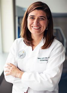
Valeria Canto-Soler, PhD
 (November 2018) Valeria Canto-Soler,
induced pluripotent stem (iPS) cells.
Human iPS cells are derived from a small, autologous, skin or blood biopsy and then coaxed, in the lab, to differentiate into a three-dimensional tissue containing all the different cell types that form the human retina properly organized into the different retinal layers. Importantly, the photoreceptor cells within these stem cell-derived human retinas have the capacity to respond to light, as photoreceptors in the normal human retina do.
“My lab brings in the expertise and technology of making the human retinal tissue that we can use for modeling disease or for developing a retinal transplant, which is really our immediate goal for CellSight as a whole,” said Canto-Soler, the Doni Solich Family Endowed Chair in Ocular Stem Cell Research, associate professor of ophthalmology, and director of the 3DRet Laboratory.
The 3DRet Lab focuses on leveraging the unique characteristics of 3D retinal organoids derived from human induced pluripotent stem cells to unveil normal and disease mechanisms involved in retinal cell specification, differentiation, maturation
Natalia Vergara, PhD
 Natalia Vergara,
“One of the things we can do with these retinas is model disease,” Vergara said. “We can model how these cells would die in situations like neurodegenerative diseases of the retina, including glaucoma, and then we can use them to screen for drugs that can protect those cells from dying.”
“There’s the potential to develop cell lines from different patients and that allows you to do something like a clinical trial in a dish,” said Vergara, who runs the Ocular Development and Translational Technologies Laboratory. “We know drugs don’t work the same in everybody, they affect people differently.”
The technology used by CellSight could make it easier to test broader samples of the population earlier in the process.
“Most of the drugs that pass preclinical studies never make it to a viable product because they fail at clinical trials,” Vergara said. “I think a lot of that has to do with fact that the models used in preclinical studies don’t really mimic what happens in a real human setting, in the population, so I think this technology really gives us the opportunity to bridge that gap.”
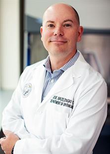
Joseph Brzezinski, PhD
 Joseph Brzezinski,
“My big goals have been to understand how you build a retina. What is the genetic blueprint that leads to a retina?” he said.
“I mostly focus on how transcription factors work because they’re the genes that execute the instructions by turning on and off other genes. They typically sit at the top of these cascades. We’ve been interested in how all cell types develop, but recently we’ve focused on rod and cone photoreceptor development.
“And especially cones. There’s a pretty big black box there on how they form and they’re difficult to study because they are pretty rare in the retina. They make up only about 2-4 percent of the mouse and human retina.”
Brzezinski heads the Laboratory of Developmental Genetics, which works to uncover the genetic programs that are used to build the retina during development and to apply them to generate therapeutic retinal cell types from stem cell sources.
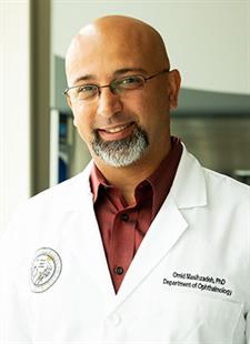
Omid Masihzadeh, PhD
 Omid Masihzadeh,
Specifically, his goal in the Laboratory of Advanced Ophthalmic Imaging is to create systems for retinal imaging that permit real-time and dynamic monitoring of living cellular processes in the retinas developed for transplanting in patients.
“The question, the billion dollar question, is what are
“The field I’m working on is called nonlinear microscopy,” Masihzadeh said. “Basically, you use such intense light – lasers – that you create nonlinearity inside the molecules of the sample. So that means the sample actually creates its own light. I shine a light with one wavelength and what comes out is completely different wavelengths, or colors. And those colors give you information about the molecules or the structure of the sample.”