Evaluation & Diagnosis
Symptoms
A variety of symptoms can occur depending on the location of the tumor. Often, tumors initially cause vague symptoms such as headache, nausea, memory impairment, or vision difficulties. Other common symptoms of skull base tumors include decreased sense of smell, hormonal abnormalities, drainage of clear fluid, nosebleeds, hearing loss, imbalance, to name a few.
Medical History & Exam
The initial evaluation consists of learning the patient’s history, or the details surrounding how the tumor became known. The duration and severity of symptoms, and the rapidity with which they developed, may be important for diagnosis or treatment planning. A physical examination is performed. This includes examination of the head and neck, and a neurological examination.
Endoscopy
Tumors of the anterior skull base share a common boundary with the sinuses. In these cases, examination of the nasal cavity with an endoscope may allow for direct visualization of the abnormality. This is a brief procedure performed in the clinic, where a nasal spray containing decongestant and lidocaine is applied to the nasal cavity, and a small (3mm) telescope is placed in the nose. If appropriate, a small biopsy can even be obtained in this fashion to diagnose a sinus tumor.
Nasal endoscopy performed in the clinic one year after surgery and postoperative radiation for treatment of a large olfactory neuroblastoma. All sinuses are patent with normal appearing mucosa, confirming that there is no evidence of disease at this point.
Imaging
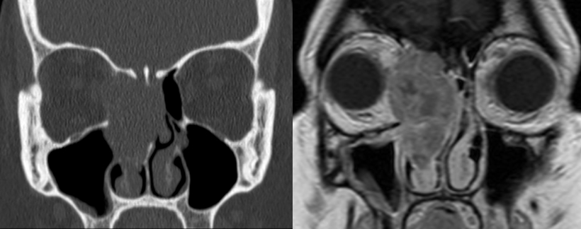
Computed tomography (CT) and magnetic resonance imaging (MRI) are the most common methods used to diagnose skull base tumors. Contrast agents may be administered by IV or orally to help outline the tumors on these types of scans. A number of other imaging modalities may be of value in specific cases.
- Magnetic resonance imaging (MRI): Magnetic fields, radio waves, and computer technology are used to create images of the soft tissues of the brain in great detail.
- Computed tomography (CT): X-rays and computer technology are used to visualize bone, collections of calcium within tumors, bleeding, and swelling.
- Functional MRI (fMRI): MRI technology is used to generate an image of brain activity according to changes of blood flow in the brain. The purpose of this test is to determine which areas of the brain control certain functions so that these areas can
be avoided during surgery.
- Positron Emission Tomography (PET): A type of scan that helps distinguish between tumor cells, necrosis (dead tumor cells), and scar tissue.
- Magnetic Resonance Angiogram/Venography (MRA or MRV): MRI technology is used to create an image of blood vessels in the brain.
- Magnetic Resonance Spectroscopy/Perfusion: These are specialized MRI-based tests that give information about the metabolic activity of specific areas of the brain, which can help in providing evidence for the presence of a tumor.

Noncontrast coronal CT (left) and T1 post-contrast MR (right) of and a squamous cell carcinoma of the right ethmoid sinus.
Biopsy
A small piece of the tumor is obtained for the neuropathologist to examine under the microscope to make the diagnosis. This procedure is called a biopsy. If a portion of the tumor is easily accessible, perhaps on nasal endoscopic examination, a simple procedure may be performed in the clinic. Often, the tumor is not easily accessible, and the biopsy is obtained and sent at the time of surgery. During surgery, the neuropathologist may be able to offer some insight on a quick initial glance, in what is called a “frozen section” analysis. Often, a number of special tests are performed on the tumor before a conclusive diagnosis is established.
Audiometry
Skull base tumors that originate near the ear or nerves that supply the inner ear often cause problems with hearing and balance function. Surgery or radiation to this area, and certain types of chemotherapy, may also affect these nerves and organs. A hearing test (audiogram) and balance testing may be recommended to assess function either before or after treatment. In this case, and audiologist will perform the necessary testing, and rehabilitation can be initiated.