Structural Heart Disease Modeling Project
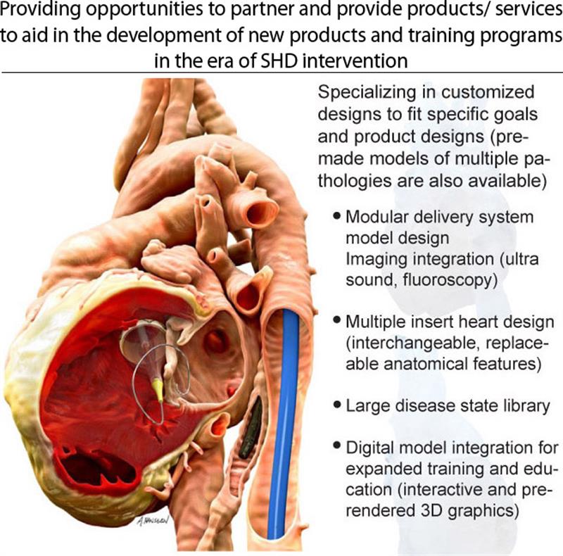
The 3D Interventional Cardiology lab is a multidisciplinary research and clinical group with a unique flow of expertise and ideas between imaging research and clinical experience. This exchange of ideas has yielded innovative research and applications for over a decade. During that time, the 3D Lab has worked with industry on a wide range of projects analyzing morphological characteristics of coronary and peripheral vasculature, worked on advanced imaging projects, and over the last four years have moved into the SHD arena with the same combination of clinical and imaging research expertise. The focus of the SHD physical model project has been a combination of clinical utility, device testing and training, and education. The most wide reaching educational component is the yearly display of our model library as part of the “Hands on Hearts” exhibit at TCT.
Through multiple internal and external projects, the 3D Lab has developed the capability to partner with companies and provide their engineering and training departments with the following services and products:
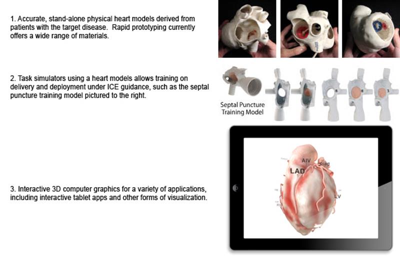
Integration of Physical Models for Multiple Applications
By combining reconstructions from both Cardiac CT and angiographic vascular models (reconstructed from two angle separated angiographic images), comprehensive and modular models can be created and customized for either a specific need or designed to be as versatile and expandable as possible. The heart design pictured below features removable aortic valve and apex insert parts. Insert parts are held in place by a combination of press-fit design, magnets and latching clips, with special care taken to make sure the part functions for the intended use with minimal or no changes to the underlying anatomical structures. Using arterial and venous reconstructions allow for realistic pathways while introducing devices. The modular nature of the vascular components allows different shapes and conditions to be added into the system at multiple locations, allowing the system to be highly versatile and expandable.
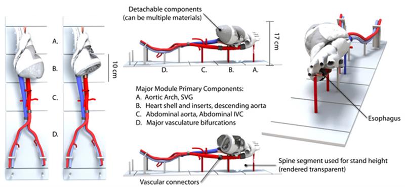
Training possibilities could be further expanded by implanting small CMOS cameras into a several optimal locations to allow direct visualization of internal structures without drastically altering the shape of the heart model (as an example, viewing windows and cut-away sections do not allow for easy direct visualization of the LAA ostium when the heart is mounted in a prone position). The ability to directly visualize anatomical features in conjunction with live imaging would give operators a unique and reinforcing understanding of catheter and device manipulation and the relationship between those manipulations, the spatial relationships and how the combination is represented in imaging.
Multiple Insert - Multi-Material Physical Models
The following computer renderings show details of a multiple insert heart model, illustrating different configurations and removable components which greatly expand the utility of a single heart model (the components pictured in yellow represent parts that are prototyped in a flexible, tissue like material). This particular design focuses on the atrial septal wall and the left atrial appendage. Starting from the far left, the heart is shown with a detachable RV free wall and all insert components removed (a) The following image (b) displays the view through a right-sided viewing window for left atrial appendage inserts. The last image (c) is a photograph of the physical model.
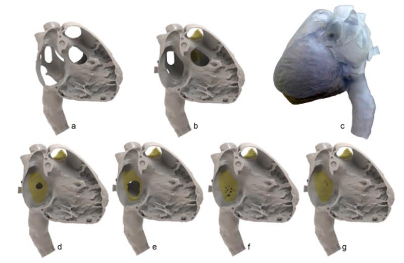
Above: four removable atrial septal inserts, starting with a small ASD (d), large ASD (e), a fenestrated ASD (f) and a PFO (g). while most inserts are generated from existing CT cases, customized inserts for specific needs can also be created.
Below: multiple endoscopic renderings of LAA ostia (blue outline), pulmonary veins (red outline) and the mitral valve (removed, purple outline), illustrating the wide variety of often non-circular shapes.

Below: multiple endoscopic renderings of LAA ostia (blue outline), pulmonary veins (red outline) and the mitral valve (removed, purple outline), illustrating the wide variety of often non-circular shapes.
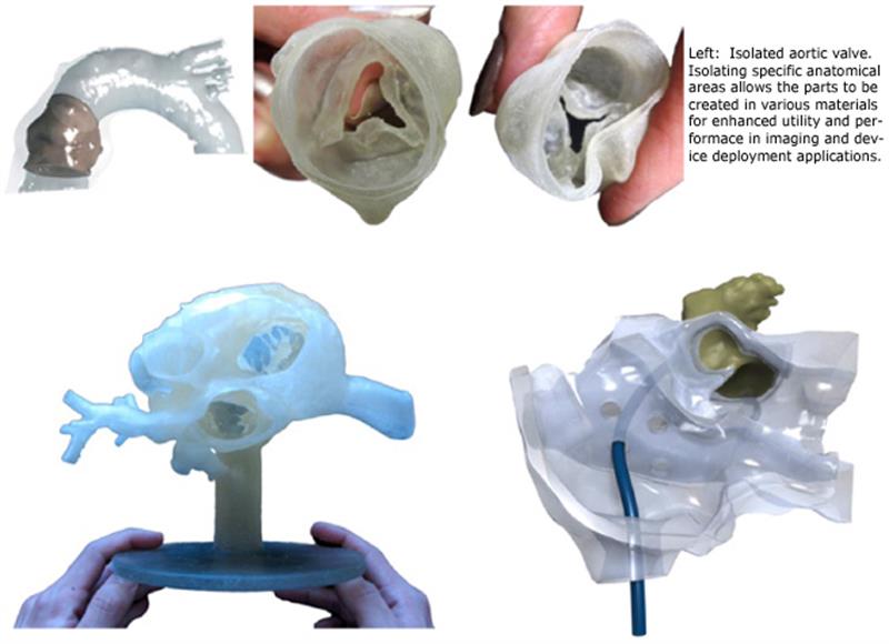
Above (left to right): smaller scale testing and training model configurations are tailored to specific needs, including different methods of mounting, additional vasculature, or highly truncated and less detailed models to focus strictly on spatial relationships
Below(left to right): smaller scale testing and training model configurations are tailored to specific needs, including different methods of mounting, additional vasculature, or highly truncated and less detailed models to focus strictly on spatial relationships between structures.
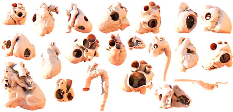
Large Disease State Library
A wide variety of pathologies have been created to aid in clinical planning which have the added benefit of a clinical interventional history to complement the physical and digital models. This collection of models represents a combination of both frequent or common cases along with some of the more unusual or complicated anatomies and cases that come through the interventional lab.
Digital Model Applications
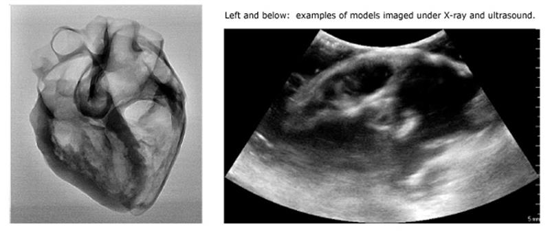
As a result of transforming volumetric images into surface mesh models, the entire library of models generated for rapid prototyping are available to be used in a variety of digital applications. Hearts or region specific sub-sets can be optimized to standard engineering and computer graphic formats, the images and the models themselves can be used to simulate different forms of imaging and the models can be integrated into educational tools.
Interactive Apps With Integrated 3D Models
Optimization of 3D Models, Warping and Deformation
Different applications have different requirements and therefore differing optimizations requirements. Models optimized for rapid prototyping differ from those optimized for use in 3D PDF documents, while “digitally grafting” anatomical features from one model to another benefit from warping the geometry from one model to another (which also offers interesting possibilities in deforming models).
Using warping techniques, the existing library of digital models allows different disease states to be “grafted” onto a single initial model. This process allows for visual and interactive representations of not only 4D characteristics but also for the chamber remodeling that can occur. The image below and to the left shows a heart with no chamber enlargement compared with right side dilation (ASD) and LV dilation (Aortic Stenosis).
