What Coronary Artery Disease Looks Like
Coronary Artery Disease Facts (US)
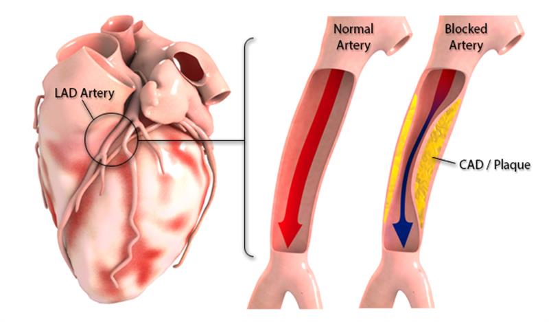
Coronary artery disease (CAD) diagnoses: 16,300,000 people (AHA 2012 Statistical Update)
Contribution to hospital stay: 1.2 million
Cost: $44 billion in expenses (The most expensive condition treated)
More than half of the hospital stays for CAD were among patients who also received coronary artery bypass graft (CABG) or percutaneous coronary intervention (PCI) during their stay.
Treatment Strategies:
- Coronary Bypass Graft (CABG, open heart surgery)
- Minimally Invasive Percutaneous Coronary Intervention (PCI)
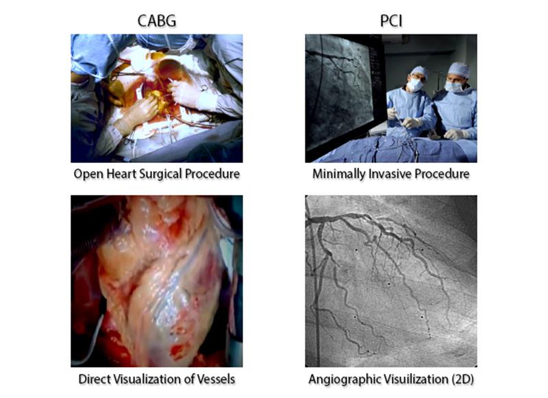
Percutaneous Coronary Intervention (PCI)
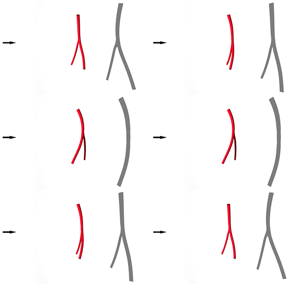
The primary 2D image interpretation challenges faced by the interventional cardiologist:
- Vessel Overlap and Vessel Foreshortening, illustrated in the simple cartoon renderings below, with the black arrows representing the X-ray source, the red vessels representing the true 3D shape and lastly the resulting 2D projection image.
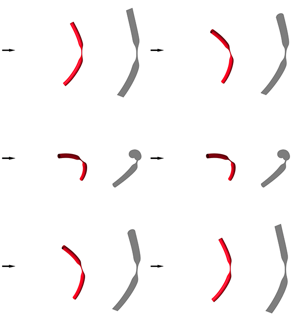
In the angiographic example below, two images of the same artery, focusing on the same arterial segment, can potentially give misleading information of blockage length and severity.
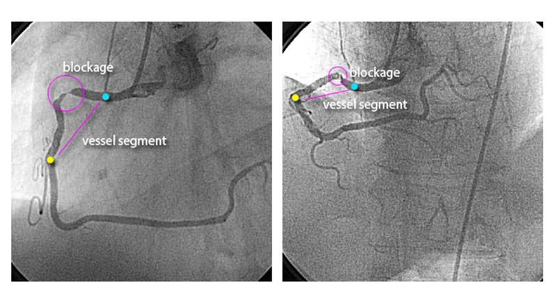
Why This Matters
Interventionalists must match pre-existing stent sizes to an estimate of the lesion length and diameter as interpreted from the images acquired, as illustrated in the following cartoon of a balloon expandable stent.
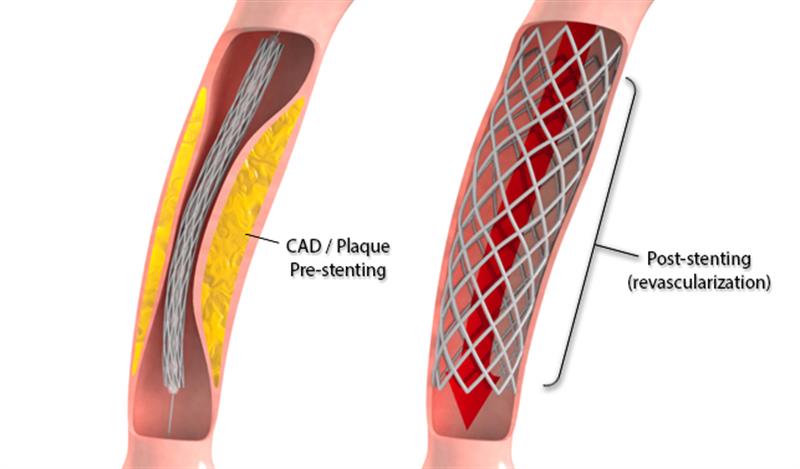
What This Means
- Stent diameter too large -->TEAR OF VESSEL WALL
- Stent diameter too small --> BLOOD CLOTS
- Stent length too short --> ADDITIONAL STENT
- Stent too long --> OTHER ARTERIES BLOCKED
- Stent in wrong location --> ANOTHER STENT
These imaging based mistakes may lead to repeat procedures and serious clinical complications.
Traditional techniques to minimize complications arising from vessel overlap and foreshortening involves taking additional X-ray pictures to ensure a complete understanding of the anatomy, which leads to:
- More radiation exposure
- More contrast dye injected
- Procedure time increased