PET/SPECT/CT
Our equipment can only accommodate small rodents and our imaging services are for research purposes only. Unfortunately, we cannot perform imaging on companion animals, such as cats and dogs.
The Animal Imaging and Irradiation Shared Resource (AIISR) performs microPET/CT and microSPECT/CT protocol development, image acquisition and image analysis for small animal models and imaging phantoms.
Nuclear medicine images are produced by giving the animal short-lived radioactive isotopes and detecting their decay using a gamma camera (SPECT) or positron emission (PET) scanner, revealing the spatial and temporal distribution of target-specific radiotracers and pharmaceuticals. An extensive array of radiopharmaceuticals, or molecular probes exist (based on 11C, 13N, 15O, 18F, 124I, 64Cu, 68Ga, 89Zr for PET and 67Ga, 123I, 99mTc, 201Ti, 111In for SPECT) to image diverse aspects of tumor physiology and biology. Data can reveal properties such as glucose metabolism, blood volume and flow, tissue uptake, receptor binding, and oxygen utilization. Since both modalities have a relatively low spatial resolution, CT is usually added for an anatomical overlay of the biodistribution of the radiolabeled probe.

Facility:
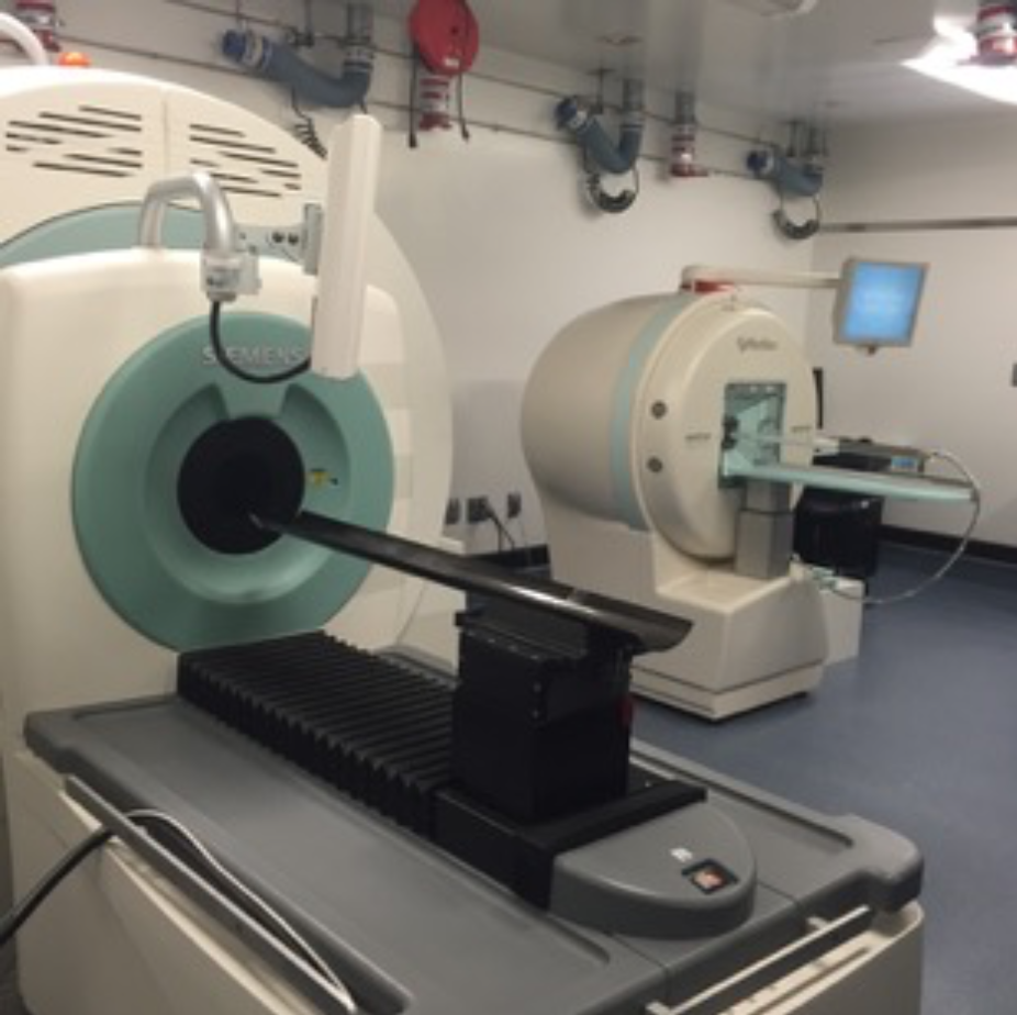
The Siemens Inveon microPET and microCT scanners were installed in 2008.
The Mediso SPECT/CT was installed in 2016.
Protocols:
- Glucose uptake and metabolism using 18FDG-PET/CT
- Dynamic and static PET/CT using novel 18F-amino acid and 11C-acetate metabolic tracers
To Schedule a Service:
Small Animal Imaging PET/SPECT/CT
- To schedule a consultation for small animal PET or SPECT studies and study design please contact Natalie Serkova, PhD.
- To schedule an imaging appointment, please reach out to the AIISR.
You must have an approved IACUC protocol. Please contact the AIISR for the instructions, SOPs, and pre-approval of PET and SPECT studies to your IACUC protocol. Please visit the Institutional Animal Care and Use Committee for more information.
PET/CT Scientific Highlights:
The University of Colorado Anschutz Medical Campus has an onsite commercial PharmaLogic Radio pharmacy facility with two GE cyclotrons 18-F and 11-C tracers) and Ga-68 generator. We also have ongoing collaborations with other academic labs for 89-Zr antibodies.

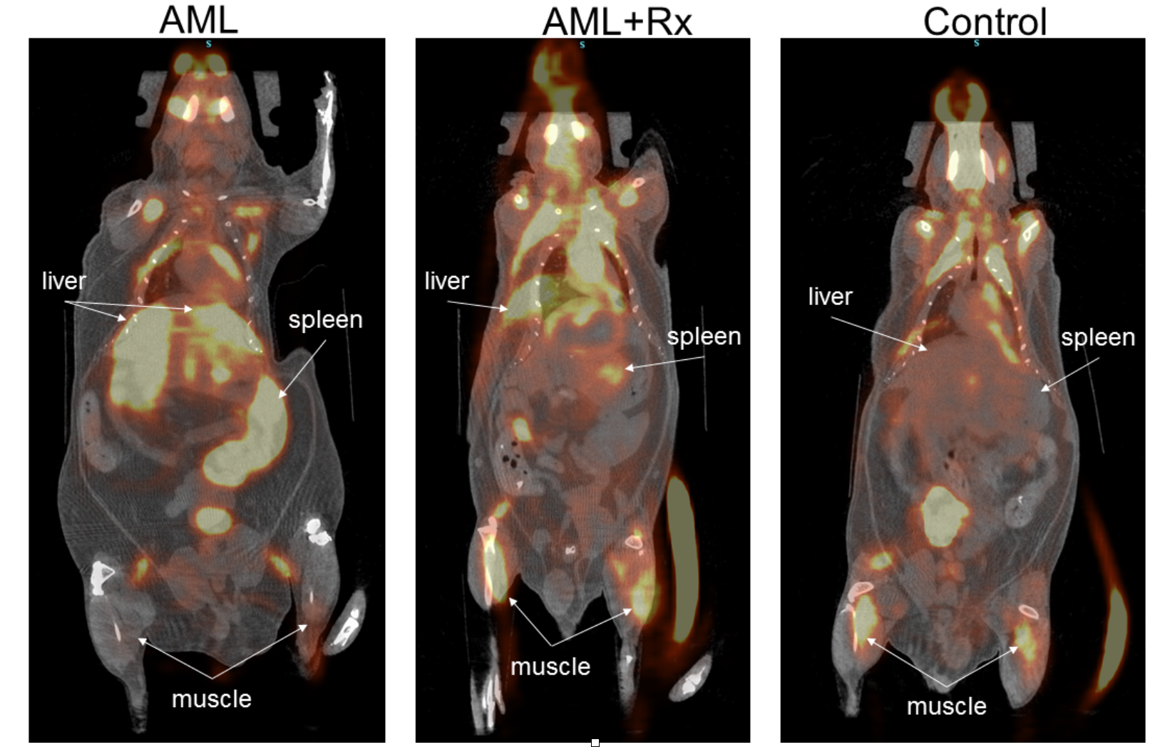
FDG-PET/CT on leukemic mice, PI: Craig Jordan, PhD (available at Cancer Cell 2018, PMID: 30270124)

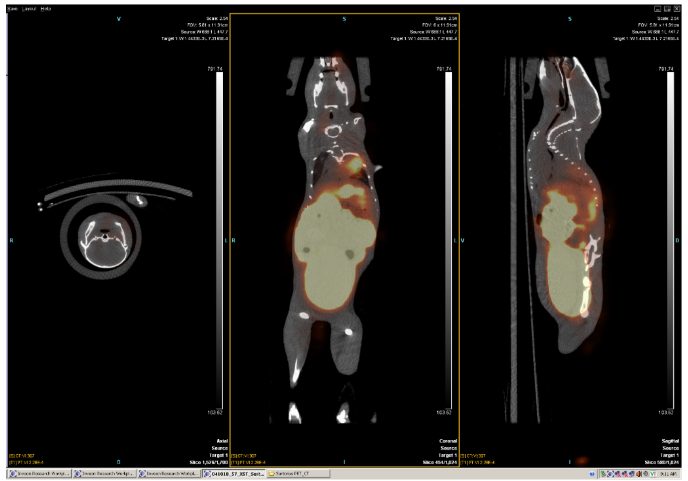
FES-PET/CT on Lung Metastases from ER+ breast cancer, PI: Carol Sartorius, PhD and Natalie Serkova, PhD
Contact Us
Natalie Serkova, PhD
Core Director
David Westerly, PhD
Core Co-Director
Tyler Weiskopf, BA
Core Manager
Vadym Zaberezhnyy, MSc
Core Technician
Qing Yuan, PhD
MR Physicist
Two more staff coming!
Location: RC1N Vivarium, Suite 1, Rm 0434