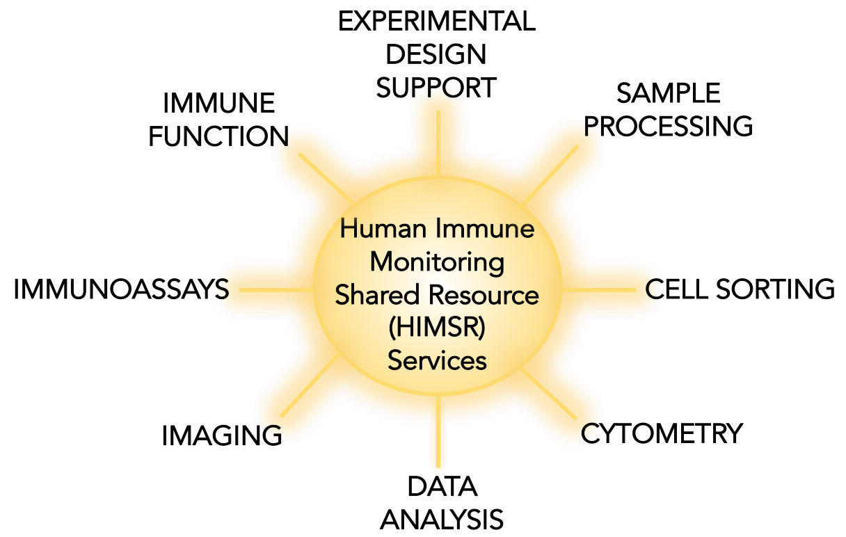
Human Immune Monitoring Services
Services
At HIMSR we help researchers fill-in gaps between clinical and basic science research, providing sample preparation and immune-based assays that fit the needs of our campus and scientific community. We also integrate with other existing core services. Contact us to tell us what you might need from us to inform our ongoing assay development process.
To better serve our campus, we offer free initial experimental design support. This includes development of research endpoints and selection of appropriate assays. It also includes testing and development of new technologies for immune monitoring.
Standard services: Available on a fee-for-service basis

SAMPLE PROCESSING
IMAGING
FLOW CYTOMETRY
IMMUNOASSAYS
DATA ANALYSIS
Customized projects: Fee-for-service quotes are available. However, we can also discuss percent effort contributions for projects involving ongoing experimental design support, assay development, or extensive analyses.
Users outside the CU system: Please inquire about external-academic rates and non-academic pricing.
Contact us: Please see iLabs or contact Angie Minic, Project Manager for HIMSR, for help building a budget that includes HIMSR services in your grant applications. The Human Immunology and Immunotherapy Initiative supported by the Dean’s Transformational Awards subsidizes CU researchers. University of Colorado Cancer Center subsidizes members' prices.