IMAGING
Identify and localize immune cells in the microenvironment of your tissue samples
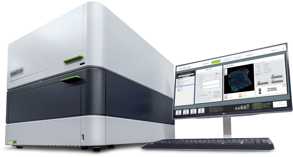
Are you interested in quantitating immune cells that infiltrate the human tissues you study, understanding where they are located, what genes they are expressing, or determining which other cells they are near in the tissue architecture? HIMSR operates a state-of-the-art spatial biology facility for many of your imaging needs. We are equipped with Akoya Biosciences’ PhenoImager HT (Vectra Polaris), the Lunaphore Comet, the 10x Genomics Xenium, and the Nanostring GeoMx.
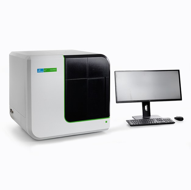
PhenoImager HT (Vectra Polaris)
The Akoya PhenoImager is a high-throughput microscope that images brightfield or immunofluorescence slides and distinguishes 7 colors in a full-slide scan or 9 colors in regions-of-interest using up to 40x objective. The antibodies are resolved with Akoya Opal technology, which allows use of the best primary antibodies for immunohistochemistry, regardless of species with no cross-reactivity.
This technology is optimal for analysis of FFPE tissues or tissue microarrays (not frozen tissues). We have 10 standardized panels to choose from that are free of development charge (see list below). Additionally, we can custom build antibody panels from over 350 different human and mouse antibodies. With each panel, we include stained tonsil-tissue controls, scan slides with up to 40x objective, and provide analysis depending on your needs.
| Pre-designed 7-color panels | Pre-designed 9-color panels |
| Basic Phenotyping Panel 1 | Basic Phenotyping Panel |
| Basic Phenotyping Panel 2 | Myeloid Panel |
| Myeloid Panel 1 | Immune Checkpoint Panel 1 |
| Myeloid Panel 2 | B-cell Panel |
| Structural Myeloid Panel | Immune Checkpoint Panel 2 |
Custom IF antibody panel
If your antibody of choice is not yet available, we can work with you to develop a custom antibody panel either in your lab or HIMSR. In the spirit of fostering collaborative research at CU|AMC, antibody panels developed in HIMSR will be shared with the campus as future standard panels.
If your antibody of choice is not yet available, we can work with you to develop a custom antibody panel either in your lab or HIMSR. In the spirit of fostering collaborative research at CU|AMC, antibody panels developed in HIMSR will be shared with the campus as future standard panels.
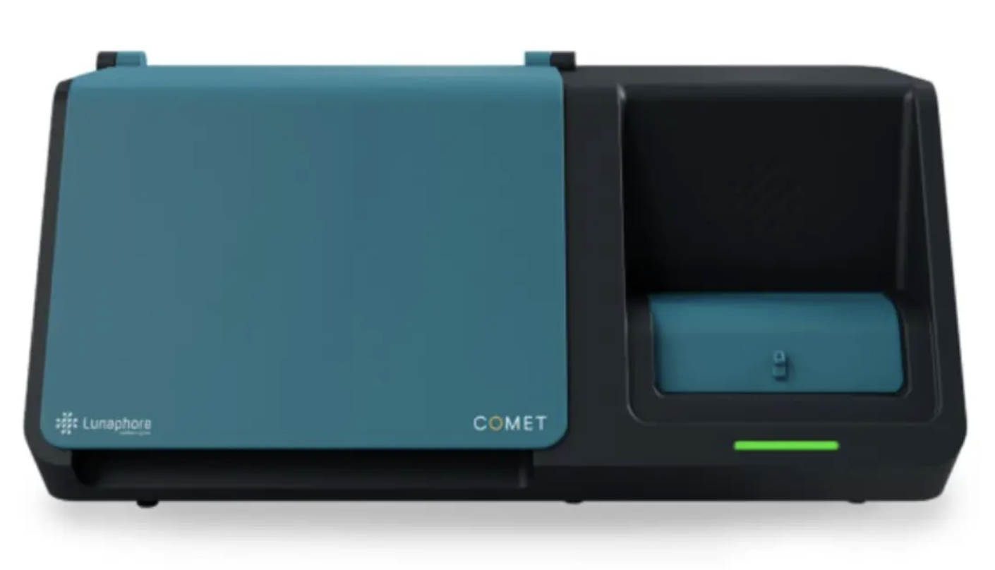
Bio-Techne Lunaphore COMET
Have you ever wished that you could have just one (or a few) more
marker(s) than what is possible on the PhenoImager? Then Lunaphore
technology is for you! This technology uses sequential cycles of
staining, imaging, and eluting so you can easily analyzed up to 40 antibody markers or 20 antibody markers and 12 RNA markers. This technology is good on FFPE or frozen tissues. Publication of data generated on this technology needs to acknowledge the shared instrument grant S10OD036353 from the NIH.
| Pre-designed Lunaphore | COMET Staining Modules |
| Module 1 Lymphocytic Markers | CD20, CD25, CD3, CD45RO, CD8, Foxp3, CD66b, CD27, CD4, CD56 |
| Module 2a Myeloid Panel 1 | CD163, CD68, DC-SIGN, HLA-DR, CD11c, CD14 |
| Module 2b Myeloid Panel 2 | CD16, CD11b, CSF-1-R, iNOS, MARCO, MERTK, pStat1, XCR1 |
| Module 3 Microenvironment and Structural Markers | HLAC1, Podoplanin (D2-40), SMA, Col3A1, CD31, Vimentin |
| Module 5 Immune State Activation Panel | CD80, PD1, Granzyme B, CD86, Lag3, Tim3 |
| Module 6 B cell | CD95/Fas, CD19, CD38, CD23, IgD, IgA, IgG, IgM |
10x Genomics Xenium
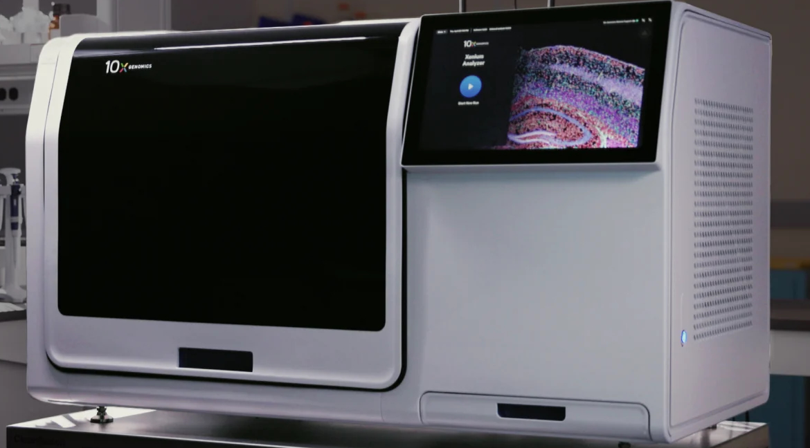
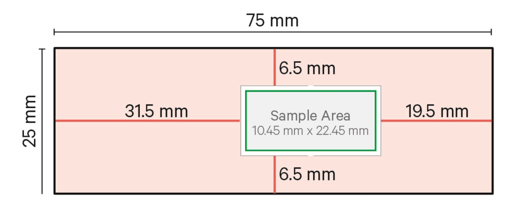
Transform your tissue samples into powerful scientific discoveries with our 10x Genomics Xenium service. We can image FFPE or fresh frozen sections on Xenium slides, using custom probes (any species) or pre-designed panels to target up to ~5,000 unique human or mice genes. Technology, including padlock probe and rolling circle amplification, ensures specific vibrant visualization. Once your data are ready, you can do analysis using Xenium Explorer to unlock insights that lead to high-impact publications and successful grant applications.
GeoMx Digital Spatial Profiler
In collaboration with the Genomics Shared Resource, we can also a spatial multiomics technology that profiles tissues for mRNA and protein expression using oligonucleotide barcoding for the whole genome.
Tissue RNA quality control
RNAscope assay used to assess RNA quality for spatial transcriptomics.
Contact
Angie Minic, MS
Project Manager
[email protected]
SAMPLE DROP BOX LOCATION
8th Floor RC1N - P18-8102
Lab phone number 303-724-3521
HIMSR SERVICE CONSULTATION REQUEST
GRANT SUPPORT DOCUMENT REQUEST