Imaging and Radiation Oncology Physics Research - Dr. Cem Altunbas
October 10 2025
We presented two studies at the ASTRO 2025 Annual Meeting in San Francisco, CA!
🎤 Rupesh’s talk: “The effect of a novel quantitative CBCT imaging method on deep learning–based image segmentation performance”
🎤 Uttam’s talk: “Improving robustness of CBCT radiomics with 2D anti-scatter grid–based CBCT”
These studies build on our ongoing work in quantitative CBCT imaging and demonstrate that quantitative CBCT can improve radiomic feature accuracy and enhance AI-based anatomical segmentation. Such advances are key to reliable CBCT-based radiotherapy outcome assessments and represent important steps toward more precise, adaptive, and data-driven cancer treatments.

July 31 2025
Memories from the 2025 AAPM Annual Meeting in Washington, DC (with Farhang, Uttam, and Rupesh). We reported on our work in four presentations.


June 15 2025
Congratulations Uttam Pyakurel! He is starting as a medical physics resident in the Department of Radiation Oncology at UCLA in July 2025!

June 1 2025
We will have several presentations at the 2025 AAPM Annual Meeting in Washington, DC, July 27-30 2025
Uttam Pyakurel:
- Evaluation of Radiomic Feature Robustness in Quantitative CBCT Imaging for Radiotherapy of Prostate Cancer
- The Effect of 2D Antiscatter Grid-Based CBCT on Tissue Visualization in the Prostate Region: An Observer Study on Tissue Delineation Accuracy
Rupesh Dotel:
- Advancing Deep Segmentation Accuracy in CBCT for Radiotherapy Via Robust Scatter Mitigation: First Results from a Pilot Trial
Farhang Bayat:
- An Adaptive Non-Local Means Filtering Method for Denoising CBCT Images Under Low X-ray Fluence Conditions
May 29 2025
Our article on improving Hounsfield Unit (HU) accuracy in dental CBCT was published in Oral Radiology. Briefly, this work shows that HU accuracy in dental CBCT images may approach the HU accuracy of gold standard diagnostic CT images by implementing a robust scatter mitigation strategy.
"Improving Hounsfield Unit accuracy in dental CBCT through implementation of 2D anti-scatter grid"

May 15 2025
Congratulations to Ryan Sabounchi! Ryan has been awarded the 2025 Graduate Education Master's Thesis Award within the University of Colorado Denver system. It is well deserved, Ryan!

March 11 2025
Our article on the feasibility of 2D antiscatter grids in clinical dental CBCT systems was published in Imaging Science in Dentistry.
February 27 2025
Our latest article investigates the implementation of our 2D antiscatter grid device in CBCT system for Proton Therapy. We would like to thank our collaborators at the University of Florida Health Proton Therapy Institute.
December 22 2024
Congratulations Rupesh Dotel! Rupesh is going to the Department of Radiation Oncology at the Baylor College of Medicine as a medical physics resident.
December 04 2024
Group brunch with Uttam, Ryan, Rupesh, and Cem. Many celebrations were long overdue.

November 20 2024
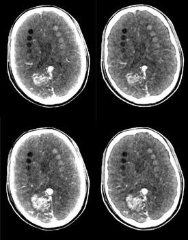
We have been exploring a compact, flat-panel detector-based CBCT approach for intracranial imaging applications. Our first proof-of-concept evaluations of this approach have just been published. You can reach the article here.
Below, the images in the left column were acquired with our in-house benchtop CBCT system, which emulates the compact CBCT geometry. The images on the right were acquired with the gold-standard MDCT. We still have some work to do in implementing several raw data correction methods, but that's something to focus on in 2025.
June 15 2024
We are wishing Farhang Bayat the best in his new endeavor: He will start his Medical Physics Residency at the Cleveland Clinic in July!
April 29 2024
Congratulations Ryan! He defended his MS thesis in Bioengineering.

December 8 2023
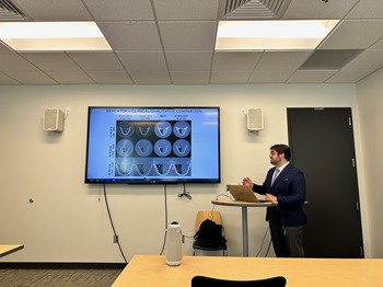
Our recent paper on quantitative CBCT imaging was selected as "Editor's Choice" in the Medical Physics Journal. You can access the article here.
December 1 2023
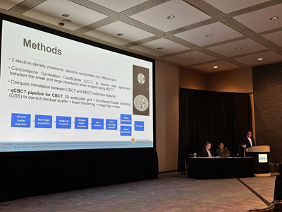
Farhang gave a presentation at the RSNA 2023 Annual Meeting about our work on radiomics feature extraction from CBCT images. His talk is titled "Improving the robustness of radiomics features extracted from CBCT images in image guided radiation therapy"
October 20 2023
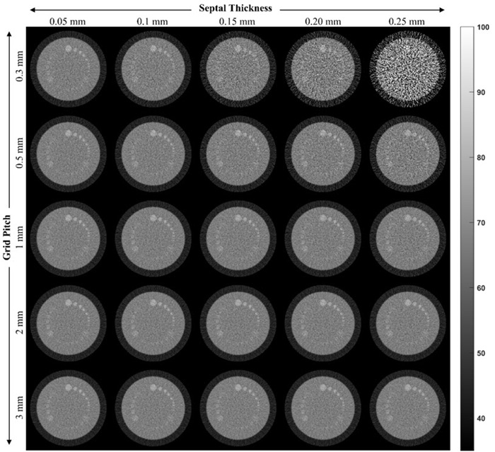
Our recent work on the simulation of 2D antiscatter grid’s primary transmission characteristics and their effect on CBCT image quality is published. Here is the link.
Besides studying the effects of primary transmission, we introduced two new methods in this work: 1) We estimate imaging system’s line spread function, LSF, (the function that characterizes the blurring introduced by the imaging chain) from 2D grid’s septal shadows. Our method showed comparable performance with respect to well-known methods based on slanted radio-opaque edge. Our approach may negate the need for additional measurements using a radio-opaque edge. 2) We developed a new method to characterize the shape of the 2D antiscatter grid’s septal shadows. The pixel size of a typical flat panel detector is too large, and hence, septal shadows are typically undersampled in an x-ray projection. Our method, called Oversampled Shadow Profile (OSP), helps to accurately characterize the shape of septal shadows in x-ray images.
The image below shows how the grid pitch and apparent septal thickness affect primary transmission and low contrast visualization in simulated CBCT images.
October 9 2023
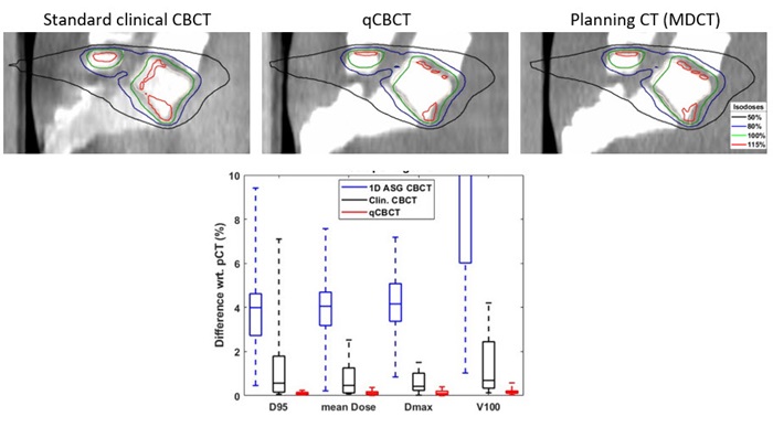
One of the dose delivery monitoring methods during radiotherapy sessions is to use cone-beam CT (CBCT) images acquired before radiotherapy delivery. Tissue densities are extracted from the images and subsequently used for dose calculations. However, density might be incorrectly represented in CBCT images, making this approach less straightforward.
Our recent preprint shows how we can directly calculate the delivered dose in CBCT images. The dose calculation accuracy of our approach was within 1% of the planning CT (considered the gold standard) in all 20 radiation treatment scenarios we evaluated. This is due to our robust scatter suppression approach in CBCT, using our 2D antiscatter grid and scatter sampling-based scatter correction in tandem.
We often receive the following question: why not just correct scatter, why use an antiscatter grid? The answer is twofold: 1) Scatter correction corrects the bias in the image signal, not the additional noise coming from scattered X-rays. Without an antiscatter grid, scatter fluence is much higher, and noise is amplified even more after scatter correction. This issue increases the reliance on noise suppression methods in CBCT images. 2) None of the scatter correction methods are perfect; there is always some error associated with scatter intensity estimation. In the human torso, scatter intensity can be so large that even a 5% error in scatter estimation can have detrimental effects on density estimation accuracy. Thus, when the scatter is reduced with a grid from the beginning, it makes scatter correction much easier.
September 5 2023
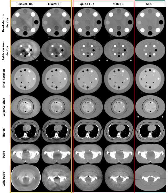
Where does the performance of our cone beam CT imaging techniques (which we develop in the lab) stand in comparison to the state-of-the-art (used in a radiation oncology clinic)? Farhang Bayat has a new publication on this subject. Here is the link: https://doi.org/10.1002/mp.16681
When conducting such evaluations, we often cut and dice the data in many ways, plotting different performance metrics. But, for experienced eyes, a picture is often worth a thousand words.
Below, the test object images within the yellow rectangle were acquired using the clinical CBCT system utilized in our clinic. The ones within the red rectangle were acquired using our techniques. Additionally, the images within the green rectangle were obtained from the helical CT scanner in our clinic, considered the gold standard. What differences can you observe among these three groups? (By the way, my daughter believes I have the “weirdest” job, as I constantly stare at circles on the computer. I suppose she has a point).
We must emphasize that a better looking image (a term inherently vague) does not necessarily correlate with improved clinical utility or impact. To provide a simplified example: an image that clearly displays soft tissues might not be the most effective image for detecting bone fractures.
August 16 2023
Going through some of our old videos: Testing the 3-axis motion stage, which holds our x-ray detector. Below: Ryan is working on the motion stage assembly.
August 5 2023
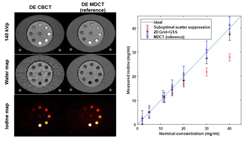
Dual energy (DE) material decomposition in CT imaging refers to splitting a CT image into 2 or 3 different material components that makes up the imaged object. For example, if a patient gets iodine contrast agent injection during a CT scan, this CT image can be decomposed into a patient anatomy-only and iodine-only images using DE techniques (In a standard CT image, iodine and patient anatomy are always blended).
This approach has numerous clinical applications in diagnostic CT imaging. However, its translation to cone beam CT (CBCT) applications (such as CBCT guided radiation therapy) has been challenging. This is in part due to poor image signal measurement accuracy when performing CBCT scans of human torso. Therefore, majority of DE CBCT applications and research is focused on smaller objects.
Cem has a new publication on improving image signal measurement accuracy in CBCT by using 2D antiscatter grids and grid-based scatter sampling (Here is the link to the Medical Physics article). Grid-based scatter sampling method employs 2D antiscatter grid as a micro-signal modulator to measure and remove residual scatter signal. This combined 2D grid + scatter sampling approach provides high signal measurement accuracy and enables DE CBCT imaging in human torso sized objects.
The sample DE CBCT image below shows how a pelvis-sized phantom is decomposed into water and iodine components.
July 31 2023
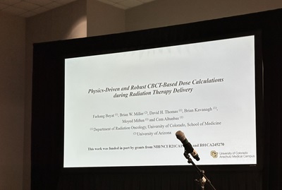
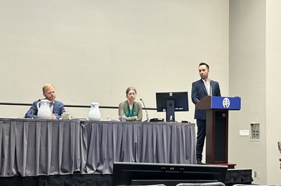
Farhang gave a presentation about his work on CBCT-based radiotherapy dose calculations at the 2023 AAPM Annual Meeting in Houston.
January 26 2023
A belated celebration of holidays and welcoming our newest team member, Uttam Pyakurel, to our group.

July 21 2022
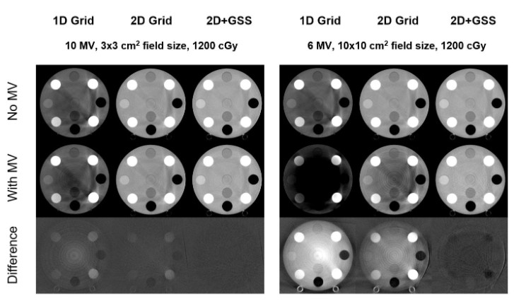
Concurrent megavoltage beam delivery and kilovoltage CBCT imaging has potential new applications in image guided radiation therapy. However, this is not a practical approach yet due to contamination of images with megavoltage cross scatter. Our recently published method addresses this challenge; it suppresses cross scatter contamination with ~300 keV mean energy, and restores image quality in kV CBCT images. Besides radiation therapy, benefits of this approach may extend to high x-ray energy nondestructive testing.
Here is the link to our paper.
Images below: When 2D antiscatter grid and gird based scatter sampling (GSS) are utilized, images without and with MV scatter contamination (1st and 2nd rows) have quite similar pixel values (indicated by the "Difference" in the third row).
July 15 2022
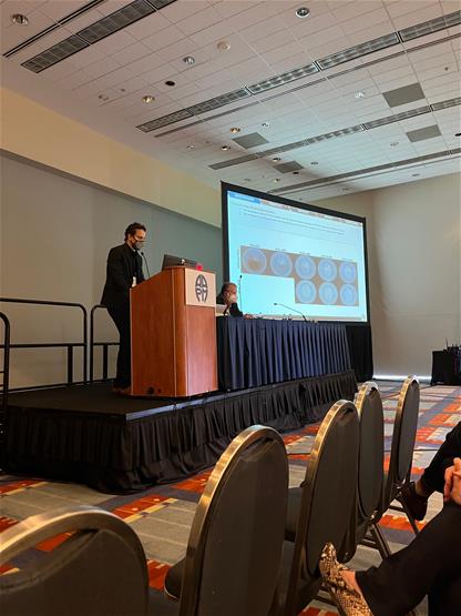
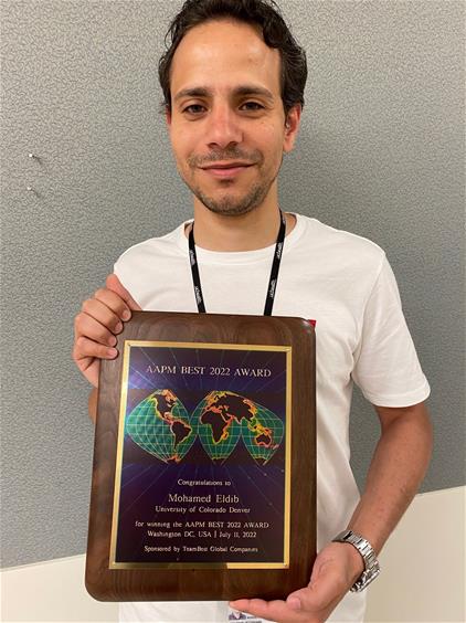
Another exciting AAPM Annual Meeting is over. Here is a picture of Eldib presenting at the CBCT session, and another one with his AAPM BEST Award.
June 1 2022
Congratulations to Mohamed Eldib, who has been selected as a recipient of the 2022 Best Medical / AAPM Fellowship.
May 20 2022
2022 AAPM Annual Meeting is around the corner, and will be held in Washington, DC. Our group will have 3 presentations on our quantitative CBCT imaging research.
- Mohamed Eldib will present our new method for quantifying the effects of CBCT gantry flex on 2D antiscatter grid performance. The link to his abstract and presentation information is here. Eldib has one more presentation on our new simulation platform to study the primary transmission characteristics of 2D antiscatter grids.
- Farhang Bayat will present our raw data correction and iterative reconstruction pipeline for CBCT-guided radiation therapy. The link to his abstract and presentation information is here.
March 5 2022
First in person meeting of 2022, SPIE Medical Imaging Conference. We presented a new approach for high fidelity kilovoltage CBCT imaging during megavoltage beam delivery in image guided radiation therapy, which was authored by Farhang Bayat.
December 10 2021
We were happy to be back in person at the RSNA 2021 Annual Meeting. We had 3 presentations. Farhang Bayat presented the effects of megavoltage cross-scatter on kilovoltage CBCT image quality in image guided radiation therapy, Mohamed Eldib presented a wavelet based image denoising method for 2D antiscatter grid CBCT, and Cem Altunbas gave a presentation on the feasibility of dual energy CBCT imaging in human torso using 2D antiscatter grids.
April 1 2021
Our work on the performance evaluation of 2D antiscatter grids and grid based scatter sampling methods was published in Medical Physics.
January 10 2021
CBCT has inferior quantitative accuracy when compared to standard multi-detector CT, also known as helical CT.
Recently, we developed a new method to improve quantitative accuracy of CBCT. This method exploits the use of 2D antiscatter grids in conjunction with flat panel detectors, to measure and correct residual scatter that was not stopped by the 2D grid. In this method, 2D antiscatter grid acts as a microarray of beam modulators placed directly on the detector, which, in turn, helps to measure residual scatter in projections. We refer this method as "Grid-based scatter sampling (GSS)". Correction of residual scatter using GSS method provides higher CT number accuracy than 2D antiscatter grid alone.
Our paper describing the GSS method was recently published in the Medical Physics Journal, and can be found here.
March 20 2020
Our work on a new scatter correction method was presented at the 2020 SPIE Medical Imaging Conference in Houston, TX. Here is recap of our presentation:
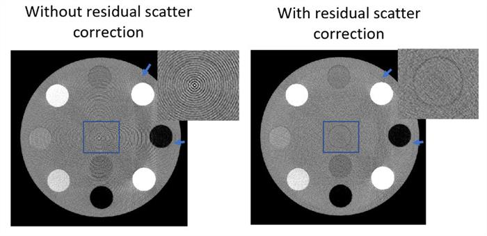
2D antiscatter grids stop vast majority of scattered radiation in flat panel detector-based CBCT. However, a small fraction of scatter goes through, and registered by the image receptor. A less known effect of residual scatter is that it may interfere with grid septal shadow correction, and cause ring artifacts in cone beam CT images.
To correct residual scatter, we developed a new method; we used 2D grid itself as a residual scatter measurement (and correction) device. This scatter correction approach not only improves CT number accuracy further, but also reduces ring artifacts.
Some of the proof of concept results can be found in the SPIE Conference Proceedings:
Click here to access the article
Here are couple of cone beam CT images from the link above. Ring artifacts are reduced substantially after residual scatter correction. Rings pointed by the blue arrows are caused by a detector related issue, not residual scatter. That’s why they are not corrected by our method.
November 15 2019
Our article, the effect of grid geometry on x-ray transmission properties, is published in Physics in Medicine and Biology.
This work shows that scattered radiation fraction is reduced by a factor of ~40 in high scatter conditions, when 2D grids (grid ratios 12-16) are used in cone beam CT geometry. Another conclusion of our work: 2D grids provide a factor of 5-7 lower scatter fraction than (high grid ratio) conventional grids, while also providing 20% better primary transmission than conventional grids.
June 10 2019
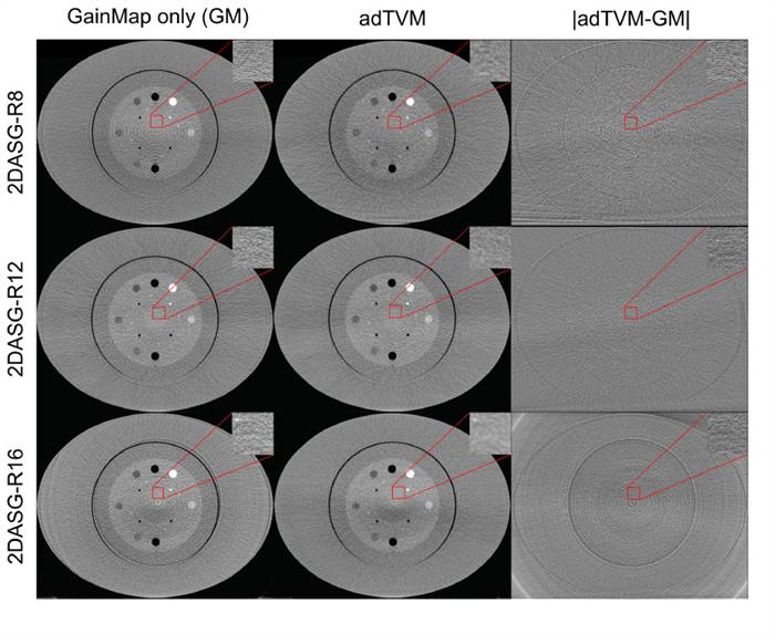
Our paper on a new ring artifact suppression method is published in the Medical Physics Journal.
In this work, we developed a new adaptive Total Variation Minimization technique (adTVM) to adjust the regularization weights in an automated fashion to reduce grid-induced ring artifacts. The images below shows the effect of adTVM method on ring artifacts in CBCT images acquired with 3 different 2D antiscatter grids.
August 10 2018
Here are a few images of 3D printed 2D antiscatter grid prototypes. These prototypes were fabricated from tungsten using Direct Metal Laser Sintering (DMLS) process. Each grid wall is only 100 microns thick, and it is aligned towards the x-ray focal spot, requiring a unique slant for each wall.
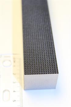
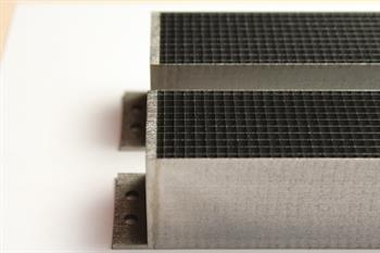
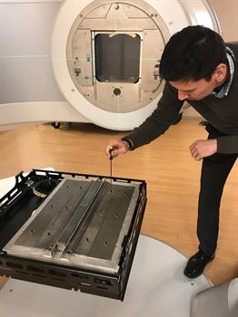
The prototype is being installed in the Varian TrueBeam CBCT system.
May 25 2018
We will have several presentations at the AAPM Annual Meeting in July. Timur’s work on CBCT ring artifacts suppression will be presented in the Science Council Session.
T. Alexeev, B. Miller, B. Kavanagh, M. Miften, C. Altunbas ”Development of High Aspect Ratio Two‐Dimensional Antiscatter Grids for CBCT”.
B. Miller, T. Alexeev, D. Thomas, K. Stuhr, B. Kavanagh, M. Miften, C. Altunbas “CBCT‐Based Dose Calculations with a Two‐Dimensional Anti‐Scatter Grid Prototype: The Effect of Scatter Suppression On Dose‐Calculation Accuracy”.
Science Council Session Presentation: T. Alexeev, B. Kavanagh, M. Miften, C. Altunbas “Development and Evaluation of a Total Variation Minimization Based Method for Suppressing Ring Artifacts in CBCT Images”.
January 20 2018
Our Medical Physics Letter is published in the Medical Physics Journal.
T. Alexeev, B. Kavanagh, M. Miften, C. Altunbas, “Two‐dimensional antiscatter grid: A novel scatter rejection device for Cone‐beam computed tomography, Med. Phys. 45(2), 529-534 (2018)
Click here to read the article.
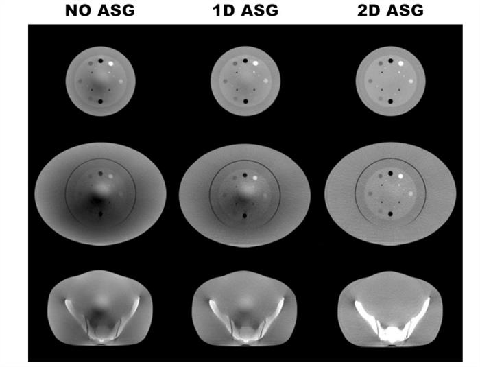
This letter reports on the first cone beam CT images acquired with our 2D antiscatter grid prototypes. We observed significantly improved Hounsfield Unit (HU) accuracy, and up to 86% improvement in contrast-to-noise ratio in images acquired with the 2D grid.
July 15 2017
We are excited that our first paper on 2D antiscatter grids for cone beam CT is published in Medical Physics Journal.
C. Altunbas, B. Kavanagh, T. Alexeev, M. Miften “Transmission characteristics of a two-dimensional antiscatter grid prototype for CBCT,” Med. Phys. 44, 3952 (2017)
You can access the free full text here.
May 29 2017
Congratulations to Timur Alexeev! His is the winner of AAPM’s Science Council Junior Investigator Competition. His work received the highest score among 170 submissions. He will give his presentation titled “Development and Evaluation of a Two-Dimensional Antiscatter Grid for CBCT” at the AAPM Annual Meeting in Denver this July.
We do not have any vacant positions at this time.
Selected publications
- Z. Yu, Y. Park, and C. Altunbas "Simultaneous scatter rejection and correction method using 2D antiscatter grids for CBCT." vol. 11312, p. 113122W. International Society for Optics and Photonics, (2020).
- C. Altunbas, T. Alexeev, M. Miften, B. Kavanagh “Effect of grid geometry on the transmission properties of 2D grids for flat detectors in CBCT,” Physics in Medicine & Biology 64, 22, 225006 (2019).
- T. Alexeev, B. Kavanagh, M. Miften, C. Altunbas “A novel total variation based ring artifact suppression method for CBCT imaging with two‐dimensional antiscatter grids,” Med. Phys. 46, 2181 (2019)
- T. Alexeev, B. Kavanagh, M. Miften, C. Altunbas “Two‐dimensional antiscatter grid: A novel scatter rejection device for Cone‐beam computed tomography,” Med. Phys. 45, 529 (2018)
- C. Altunbas, B. Kavanagh, T. Alexeev, M. Miften “Transmission characteristics of a two dimensional antiscatter grid prototype for CBCT,” Med. Phys. 44, 3952 (2017)
- C. Altunbas, Y. Zhong, C. Lai, C. Shaw, “Reduction of ring artifacts in CBCT: Detection and correction of pixel gain variations in flat panel detectors,” Med. Phys. 41, 091913 (2014)
- C. Altunbas, “Image corrections for scattered radiation” chapter in “Cone Beam Computed Tomography,” Ed. C. Shaw, Taylor&Francis, Boca Raton (2014)
- C. Altunbas, T. C. Hankinson, M. Miften, T. Tello, S. R. Plimpton, K. Stuhr, A. K. Liu,” Rotational setup errors in pediatric stereotactic radiation therapy,” Prac. Rad. Onc., 3(3),194-198 (2013)
- C. Glide-Hurst, M. Bellon, R. Foster, C. Altunbas, M. Speiser, M. Altman, D. Westerly, N. Wen, B. Zhao, M. Miften, I. J. Chetty, and T. Solberg,” Commissioning of the Varian TrueBeam linear accelerator: A multi-institutional study,” Med. Phys. 40, 031719 (2013)
- L. Chen, C. C. Shaw, C. Altunbas, C. Lai, X. Liu, ” Spatial resolution properties in cone beam CT : A simulation study,” Med. Phys. 35, 724-734 (2008)
- C. Lai, C. C. Shaw, L. Chen, C. Altunbas, X. Liu, T. Han, T. Wang, “Visibility of microcalcification in cone beam breast CT: Effects of x-ray tube voltage and radiation dose,” Med. Phys. 34, 2995-3004 (2007)
- C. Altunbas, C. C. Shaw, L. Chen, C. Lai, X. Liu, T. Han, T. Wang, “A post-reconstruction method to correct cupping artifacts in cone beam breast computed tomography,” Med. Phys. 34, 3109-3118 (2007)
- X. Liu, C. C. Shaw, T. Wang, L. Chen, C. Altunbas, S. C. Kappadath, “An accurate scatter measurement and correction technique for cone beam breast CT imaging using scanning sampled measurement (SSM) technique," Proc. SPIE, 6142, 614234 (2006)
- X. Liu, C. C. Shaw, C. Altunbas, T. Wang, “Electronic Collimation for Implementing Slot-Scan Digital Radiography with a Flat-Panel Detector – Initial Experience for Scatter Reduction in Chest Phantom Images,” IEEE Trans. Med. Imag. 25 (4), 496-502 (2006)
- C. Altunbas, E. F. Barasch, D. R. Bednarek, S. Rudin, “Photon-counting energy-sensitive micropattern gas detector for digital radiography,” Proc. SPIE 5030, 833-844 (2003)
- C. Altunbas, M. Capéans, K. Dehmelt, J. Ehlers, J. Friedrich, I. Konorov, A. Gandi, S. Kappler, B. Ketzer, R. De Oliveira et al., ”Construction, test and commissioning of the triple-gem tracking detector for COMPASS,” Nucl. Instr. Meth. A 490, 177-203 (2002)
 Cem Altunbas, PhD is an Associate Professor in the Department of Radiation Oncology at the University of Colorado School of Medicine (SOM). Dr. Altunbas’ research is focused on the development of novel x-ray imaging instruments and methods for
medical imaging and image-guided radiation therapy. Before joining University of Colorado SOM, he has investigated photon counting gaseous detectors for digital radiography at SUNY Buffalo, and helped to develop a chest radiography system for
lung nodule detection and a prototype breast computed tomography system for breast cancer imaging at MD Anderson Cancer Center. After completing his medical physics residency at Medical College of Virginia, he joined the Department of Radiation
Oncology at University of Colorado SOM, and he has been working on imaging challenges faced in cone beam computed tomography (CBCT) guided radiation therapy. Currently, his NIH funded research is centered on developing a new 3D-printed antiscatter
rid to improve soft tissue visualization and quantitative image features in CBCT.
 Ryan Sabounchi is a design engineer at Altunbas Lab and developing a benchtop CBCT system to evaluate and enhance the efficacy of 2D antiscatter grids for scatter reduction in CBCT systems. Ryan graduated with his B. Sc. degree in mechanical engineering from the University of Colorado Denver in 2019 and M.S. degree in bioengineering from the University of Colorado Anschutz Medical Campus in 2024. In his free time, Ryan enjoys video game development, weightlifting, and working on his car.

Uttam Pyakurel is a Postdoctoral Fellow in the Department of Radiation Oncology at the University of Colorado School of Medicine since January 2023. He received his Bachelor's and Master's degrees from Tribhuvan University, Kathmandu, Nepal, in 2012 and 2016, respectively. In 2022, he received his Ph.D. degree from the University at Albany, Albany, NY, with the prestigious 'Distinguished Doctoral Dissertation Award.' His Ph.D. research focused on x-ray phase imaging and its potential to improve the accuracy of breast cancer detection in mammography. Normally, phase imaging is carried out using synchrotron sources due to the requirement of high spatial coherence of the beam. However, he specifically developed a lab-based x-ray phase imaging system using a conventional source that could potentially be translated into the clinics for diagnostic imaging. Additionally, he demonstrated that the dark-field images obtained simultaneously from the same system provided stronger contrast and additional features. Currently, Uttam's research focuses on the development of quantitative cone beam CT imaging methods for image guided radiation therapy.