Cell and Tissue Analysis Core
Overview
The Cell and Tissue Analysis Core provides diabetes researchers at the University of Colorado with a unified means to access and effectively use state-of-the art microscopy and mass cytometry systems to drive innovative research. This necessarily includes providing access to this equipment; expertise and training to use the equipment effectively for diabetes-related tissues; expertise and validated diabetes- related resources to prepare samples appropriately; guidance to design experiments; resources to analyze data; and assistance with its interpretation.

Advanced Microscopy
Advanced Light microscopy core- The ALMC provides a suite of state of the art microscopy systems with broad capabilities and dedicated professional staff.
-Multi-color, high sensitivity confocal and two-photon microscopy
Zeiss LSM780 NLO (with 2-photon excitation and FLIM/FCS)
Olympus FV1000 (with CARS/SRS)
3I Marianas inverted spinning disk microscope
3I Vivo upright spinning disk microscope (with 2-photon excitation)
-Super-resolution nanoscopy (STED, STORM/PALM)
Zeiss Elyria TIRF/STORM
Home-built STED system (2-color)
-Fluorescence lifetime imaging (metabolism, oxidized lipids, FRET molecular interactions)
-Coherent Raman imaging (CARS/SRS lipid imaging)
-Subsidized services for diabetes researchers
Intra-vital microscopy-Imaging systems and expertise is available for custom intra-vital imaging for diabetes applications.
-Abdominal window preparations
-Peripheral nerve preparation
-Intravital imaging and tissue explant imaging
-Upright 2-photon systems
Leica SP8 DIVE (700-1300nm)
Olympus FV1000 with DIVER detector (ultrasensitive detection)
-Whole organ imaging with Light sheet microscope and tissue clearing (coming soon)
Reserve an ALMC instrument (DIVER and light sheet)
Request custom intra-vital imaging services
Consulting and assistance
-Expert assistance is available for diabetes researchers to incorporate new advanced microscopy approaches to enhance their research. This includes advice on experimental design; technical assistance; assistance with image quantification; and advice for sample preparation.
-We carry specialized reagents needed for certain imaging modalities (e.g. super-resolution imaging), as well as standards for system calibration (e.g. test charts, fluorophore standards).
-The ALMC seminar series introduces new advanced systems and capabilities or covers fundamental concepts in microscopy.
Cytometry
Spectral flow cytometry- Spectral flow cytometry acquires the full fluorescence spectrum, allowing ~25 channel detection with autofluorescence rejection. This can use conventional fluorescence-based antibodies, can analyze fluorescent protein reporters, and retains the speed of conventional flow analysis.
-Cytek spectral flow cytometers analyzers
3-laser Aurora (405nm, 488nm, 640nm)
5-laser Aurora (355nm, 405nm, 488nm, 561nm, 640nm)
-Advice with custom panel design
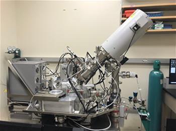
Multiplexed Ion beam Imaging- MIBI combines the multiplexing of rare-earth metal labels and time-of-flight detection, with scanning ion beam vaporization. Together, this technology enables highly multiplexed (up to 40 channel) imaging with sub-cellular (250nm) spatial resolution, detection of low abundance markers, and a large dynamic range.
-IONpath MIBIscope system (full service imaging)
-Sample preparation
-Custom antibody probe generation and validation
-Validated Diabetes specific probes (coming soon)
-Multiplexed imaging analysis (QC, segmentation, population analysis, spatial analysis).
-Consultation services
Richard Benninger, PhD
Co-director
Richard.benninger@cuanschutz.edu

Rachel Friedman, PhD
Co-director
Rachel.s.friedman@cuanschutz.edu
