Exciting Discoveries
Department of Biochemistry & Molecular Genetics
We highlight research articles on our website to keep you informed about the latest advancements and exciting discoveries from our amazing scientists within the basic science departments at CU Anschutz Medical Campus.
By showcasing our cutting-edge research, we hope to spark curiosity, promote understanding, and inspire engagement with science and innovation. Our goal is to empower you with knowledge that can inform decision-making, foster critical thinking, and contribute to a better understanding of what we do here in the research labs at the University of Colorado Anschutz.
2024

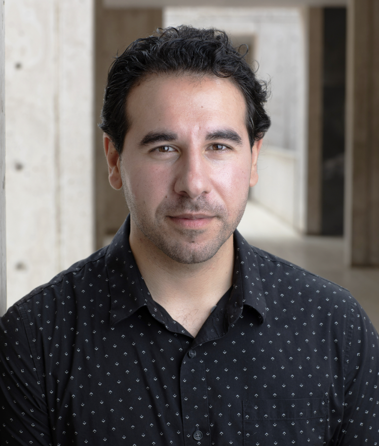
Dr. Catherine ‘Cat’ Musselman studies chemical signals that influence how DNA is packaged and used inside our cells. Our DNA is like a cook book that is filled with instructions (or recipes) to make thousands of proteins. Just like a classic cook book that contains recipes for all different types of food, such as casseroles, cheeseburgers, and cakes, our DNA cook book contains the instructions to make all different types of proteins, such as insulin (to break down sugar), collagen (to provide structure and support for skin, muscles, and bone) or hemoglobin (transports oxygen in blood). You might be interested in baking a birthday cake so you select and use a recipe for chocolate cake while leaving all of the other 100s of recipes in the cook book unused. That’s what happens inside every cell in our body every day—every cell in our body contains a full DNA cook book complete with the instructions to make every protein! Cat and her research team are dedicated to learning more about how chemical signals influence how DNA is packaged and used—turned on or off—and the role they play in the normal every day cellular functioning as well as the development of disease states such as cancer.
More information about Cat's research can be found on her lab's website here.
Dr. Cat Musselman and her team published a science article about her research in the journal of eLife in February 2024 titled, ‘Nucleosome conformation dictates the histone code.’
To gain a better understanding of the research in this research article, please read the easy-to-understand summary below.
Nucleosome conformation dictates the histone code
DNA can be thought of as a long thread that contains instructions for how your body works. To fit all this information into the center of a cell, this long thread needs to be wrapped around “spools” called histones to make it all fit inside the cell. Together, the histone “spools” and DNA “thread” form tightly wrapped bundles called nucleosomes. Each nucleosome looks like a bead on a string and their job is to help organize and control access to your DNA. Part of this organizational/control system is the placement of chemical tags on the histone “spools” that tell the cell what to do with the wrapped DNA. Like a code, some tags say, “this part of the DNA is important—read it!” (turning on genes) while other tags might say “this part of the DNA is not needed right now—ignore it.” (turning genes off).
These chemical tags are modification made above the genes or DNA, which is why they are called ‘epigenetic’ or ‘above the genes,’ and do not change the sequence of the DNA. They help the cell decide which parts of the DNA to use and when. Importantly, these tags can be misplaced—and sometimes even lost—in a variety of diseases including cancer, making it very important that we understand how they work.
The traditional understanding is that when a chemical tag is present the tag will be easily read and the gene will be turned on or off. However, this has been researched with just the histone “spool”, ignoring the thread of wrapped DNA. The research from Dr. Musselman’s team in collaboration with the company Epicypher (https://www.epicypher.com), suggests that this process is not as straightforward as originally thought and that some chemical tags may be hidden in the tightly wrapped DNA making them harder to “read”. Their research suggests that sometimes more than one chemical tag is needed, with some tags changing the nucleosome itself then allowing for proteins to recognize and read the other tags.
This research suggests that the code formed by these chemical tags is more complex than originally thought and we must include wrapped DNA in future research studies to fully understand how these tags are interpreted. This is critical information for developing and refining drugs that target the chemical tags. These types of drugs are called ‘epigenomic therapies’ because they would target the chemical tag modifications made above the DNA.
Some epigenomic therapies are already FDA approved for treatment of various diseases including blood disorders, cancer (such as acute myeloid leukemia, multiple myeloma and breast cancer), and most recently in the treatment of Duchenne muscular dystrophy. There are a large number of other diseases in which epigenomic therapies hold great promise including for autoimmune and neurological disorders. Additional studies such as this one, are critical to move forward on developing therapeutics for a wide variety of diseases.
Dr. Joe Nassour studies telomeres, the protective cap located at the ends of chromosomes. Chromosomes store our genetic instructions inside every cell in our bodies. You can think of chromosomes as the library that stores the genetic instructions to make everything needed by your body to live. Each time a cell divides and the organism ages, the telomeres (protective cap) naturally shorten, gradually losing their protective function. When telomeres become too short, cells can no longer divide, leading to the loss of function of tissues and organs. Joe and his research team are dedicated to learning more about how the length of telomeres influence the immune system and the role they may play in the development of cancer.
More information about Joe's research can be found on his lab's website here.
Dr. Joe Nassour and his team published a review science article about his research in the journal of DNA Repair in January 2024 titled, ‘Telomeres as hotspots for innate immunity and inflammation.’
To gain a better understanding of the research in this review article, please read the easy-to-understand summary below.
Telomeres as hotspots for innate immunity and inflammation
As we age, our bodies undergo gradual changes that affect how our organs work, making us more at risk for common diseases. Scientists are studying exactly how aging happens, and they've found some key factors at the cellular and molecular levels. In this review paper, Joe and his research team focused on two important factors: problems with telomeres (the protective caps on the ends of our chromosomes) and inflammation (the recruitment of immune cells to damage or infection). These factors – telomere problems and inflammation – seem to play significant roles in the aging process. The Nassour team found that when telomeres aren't properly protected and shorten, it can lead to inflammation, potentially contributing to aging and related diseases. By looking at the interaction between telomeres, our immune system, and inflammation, Joe and his team hope to better understand the aging process and why we become more susceptible to illness as we grow older.


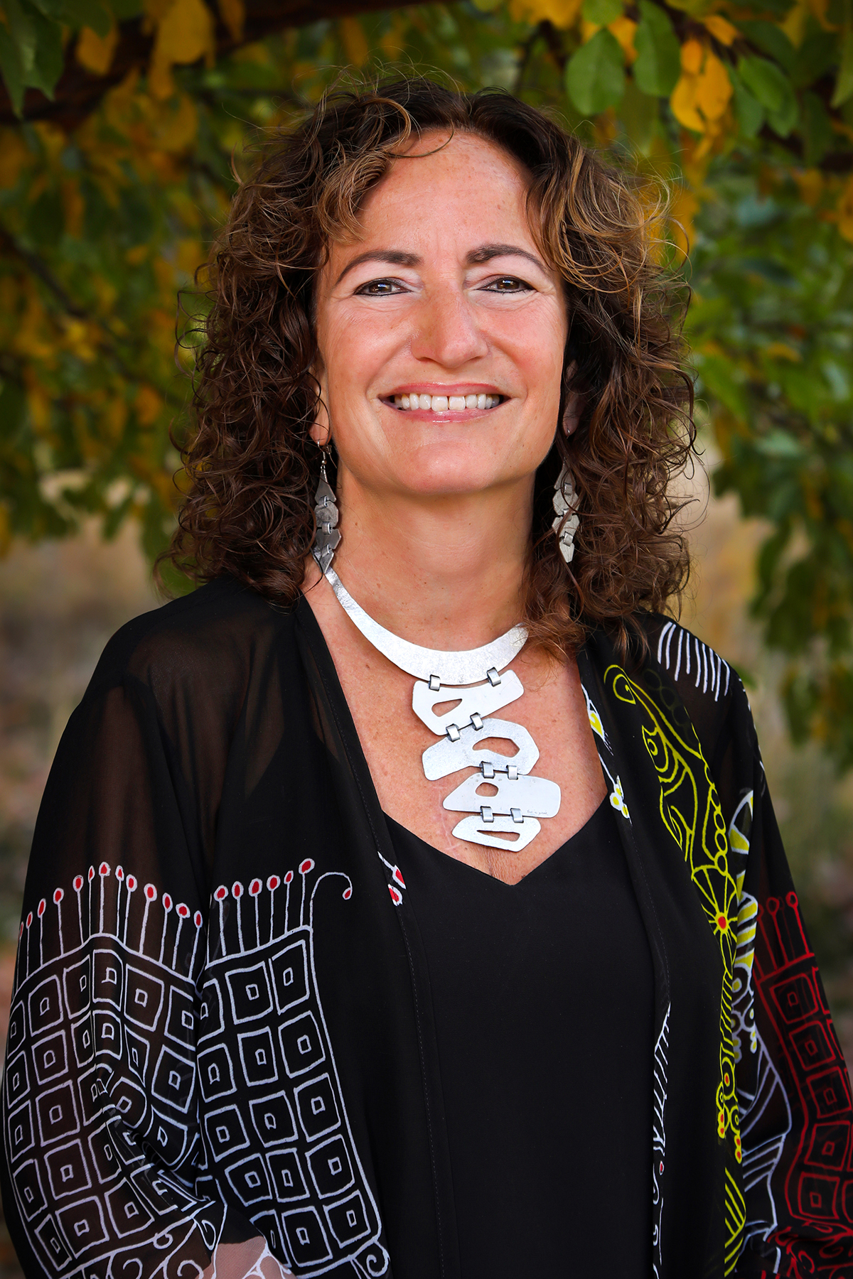
Dr. Julie Cooper studies the role of the protective caps – called telomeres – at the ends of chromosomes. Through her research she has discovered that telomeres are so much more than just a protective cap! Telomeres play important roles in the stability of the genetic instructions within chromosome as well as regulation of the cell cycle through communication with a variety of proteins. Every day Julie and her research team learn more about the many important roles carried out by telomeres!
More information about Julie’s research can be found on her profile page here.
Dr. Cooper and her team published a science article about her research in the journal of Nature Communications in 2024 titled, ‘Fate of telomere entanglements is dictated by the timing of anaphase midregion nuclear envelope breakdown.’
To gain a better understanding of the research in this article, please read the easy-to-understand summary below.
Fate of telomere entanglements is dictated by the timing of anaphase midregion nuclear envelope breakdown
Inside every cell, the genetic instructions (also called DNA) are found on what looks like extremely long pieces of yarn, tightly packaged into compact and organized bundles called chromosomes. In order to divide - or make more copies of each cell - the DNA in the tightly bundled chromosomes loosen into less organized and slightly messy piles of yarn so that every section of the DNA can be copied.
Sometimes, the copying process can get stuck, causing parts of the DNA to become tangled. If these tangles are not fixed, it can result in the death of the cell. Julie, Rishi (the first author of the paper and a post-doctoral fellow in Dr. Cooper’s lab) and their research team found a few proteins that play important roles in cell division, specifically the detangling process, in the fission yeast they study. They found a protein called Taz1 that normally helps prevent these tangles during cell division. Without Taz1, tangles form more easily, especially at colder temperatures below 20°C. They also found another protein, called Rif1, that makes it harder to untangle these knots.
During cell division, the cell's nucleus (which holds the DNA) breaks down in the middle, allowing the tangles to be fixed in the surrounding cell fluid. However, Rif1 delays this breakdown in cells without Taz1 at low temperatures, making it difficult to untangle the DNA and causing the cell to die. When there is too much Rif1, even in normal cells, it causes similar problems at low temperatures. But if the cell membrane is made more fluid, these problems can be avoided.
So, the ability for cells to copy the genetic instructions correctly without being delayed by messy, tangled bits of DNA, a variety of proteins – specifically Taz1 – must be available for cell division success!
2023

Dr. Anthony Saviola studies snake venom and how it affects the body. He uses powerful research techniques to identify and detect signals in the body – called biomarkers – that play a role in the body's response to injury from snake bites.
More information about Anthony’s research can be found on his Google Scholars page here.
Dr. Saviola and his research colleagues published a science article about his research in the journal of the Analytical Science Advances in 2023 titled, ‘Feasibility of detecting snake envenomation biomarkers from dried blood spots.’
To gain a better understanding of the research in this article, please read the easy-to-understand summary below.
Feasibility of detecting snake envenomation biomarkers from dried blood spots
Have you ever wondered how the venom from snake bites works? Why it causes so much damage and sometimes leads to the death of the person or animal that was bitten? This is the type of research that Dr. Anthony Saviola works on – figuring out how snake venom affects the body. Anthony is interested in looking for markers in the blood of people who have been bitten by snakes to find out what happens. What types of responses can be detected in the blood? What important proteins or biomolecules do snake venoms interact with? Is there anything he can use to figure out how to stop snake venom from causing harm and/or death? These are the types of questions his research seeks to answer.
So how do they do study snake venom and its effects on the body? Anthony and his research team utilized a technique – called biofluid proteomics – that analyzes small samples of blood to identify biomarkers and detect changes in response to injury from snake bites!
A biomarker is like a signal in your body that changes depending on your health status and what's happening inside your body. As an example, when you have a fever, the heat from your forehead is a biomarker that signals you might be sick. In the same way, scientists can look for these signals in your blood to understand if you're healthy or if you have a certain illness. These biomarker signals can be things like certain proteins, genes, or chemicals that change when something is wrong. So, identifying and detecting biomarkers helps scientists learn more about our response to injury and disease.
In this study, Anthony and his team used small dots of dried blood from a fatal rattlesnake bite case to try to find biomarkers linked to snake venom. They found many proteins that were either unique to or more prevalent in the envenomated blood (person bit by the rattlesnake).
2022

Dr. Lydia Heasley studies the genome (genetic instructions) in a specific type of budding yeast called Saccharomyces cerevisiae. She aims to understand how the genome changes and evolves over time and how variations in the genome influence the yeast's ability to adapt to its environment.
More information about Lydia's research can be found on her lab's website here.
Dr. Heasley and her team published a science article about her research in the journal of Frontiers in Genetics in June 2022 titled, ‘Bursts of genomic instability potentiate phenotypic and genomic diversification in Saccharomyces cerevisiae.’
To gain a better understanding of the research in this article, please read the easy-to-understand summary below.
Bursts of genomic instability potentiate phenotypic and genomic diversification in Saccharomyces cerevisiae
Humans are thinking a lot about how we might solve emerging problems due to the new pressures of climate change in a warming world, but what are microbes doing? This is a question Dr. Lydia Heasley has focused her research on by studying how Saccharomyces cerevisiae – a common yeast – is able to adapt to changes in their environment. Lydia and her research team looked at different yeast strains and saw that there were large structural changes to the way that the yeast stores its genetic information. Think of reorganizing a messy closet – you may find an old pair of shoes you want to wear again or you might remove and donate a few old pairs of jeans that you will never wear again. You might even add a newly purchased sweater and pair of socks that now need a place to be stored in the closet. Successfully removing old clothes or adding others to the closet results in organizational or ‘structural’ changes to your closet. This is similar to what happens to genetic information in yeast. These big, structural changes to the way the genetic information is stored allowed the yeast cells to exhibit new behaviors and functions, such as a change in the physical shape of the cell or an ability to resist drug treatment. The structural changes are then passed on to the next generation of yeast cells which allows for the whole population of yeast to have more diverse and flexible genetic instructions, just like humans who have different talents and abilities. As unpredictable environmental conditions arise – like a warming environment or being treated with drugs – some of the yeast cells with the most helpful structural changes are able to survive these threats, enabling them to pass on those characteristics to the next generation which helps the yeast species survive as a whole.
2019

Dr. Ning Zhao studies proteins in real time through their entire life cycle – from birth to death – inside live cells. She has developed new highly advanced ways to visually track and study proteins inside live cells. This research helps us see exactly when, where, and how proteins are made, folded, changed, and broken down in both healthy and diseased cells.
More information about Ning’s research can be found on her lab’s website here.
Dr. Zhao and her team published a science article about her research in the journal of Nature Communications in 2019 titled, ‘A genetically encoded probe for imaging nascent and mature HA-tagged proteins in vivo.’
To gain a better understanding of the research in this article, please read the easy-to-understand summary below.
A genetically encoded probe for imaging nascent and mature HA-tagged proteins in vivo
We, as humans, enjoy tracking the progress and important milestones of our children and/or loved ones over their lifetime. We take photos of celebrations and special events, like birthdays, graduations and moving into a new home or a new city. We mark the wall to monitor our children’s growth as they age each year. Some of us might even use the ‘find a friend’ app on our phones to pinpoint the location of a loved one. This is the type of research that Dr. Ning Zhao has focused her research on – tracking the progress and important milestones of proteins inside of live cells. Ning is interested in following proteins – from birth to death – inside live cells to find out what happens to the proteins over their lifespan. Where do they go? What other proteins or biomolecules do they interact with? How long do they live? What happens when they break down and die? Do proteins act differently in healthy cells compared to diseased cells? These are the types of questions her research seeks to answer.
So how do they do it? Ning and her research team developed a new and innovative imaging technique specifically for this purpose! They developed a tracking device, called a probe, that attaches to proteins in various parts of the cell and emits a color that can be seen under a microscope. The tracking device has been given the nickname ‘frankenbody’ because it consists of molecules that have been stitched together to create the probe (similar to Frankenstein). Ning and her team have found that this new probe works incredibly well and is very successful even when tracking single molecules inside living cells, which is like finding 1 individual person in a crowded city teeming with thousands of people! This kind of ‘frankenbody’ probe is a very powerful research tool that can be used to study the movement and life cycle of proteins in living cells.