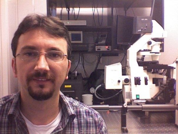
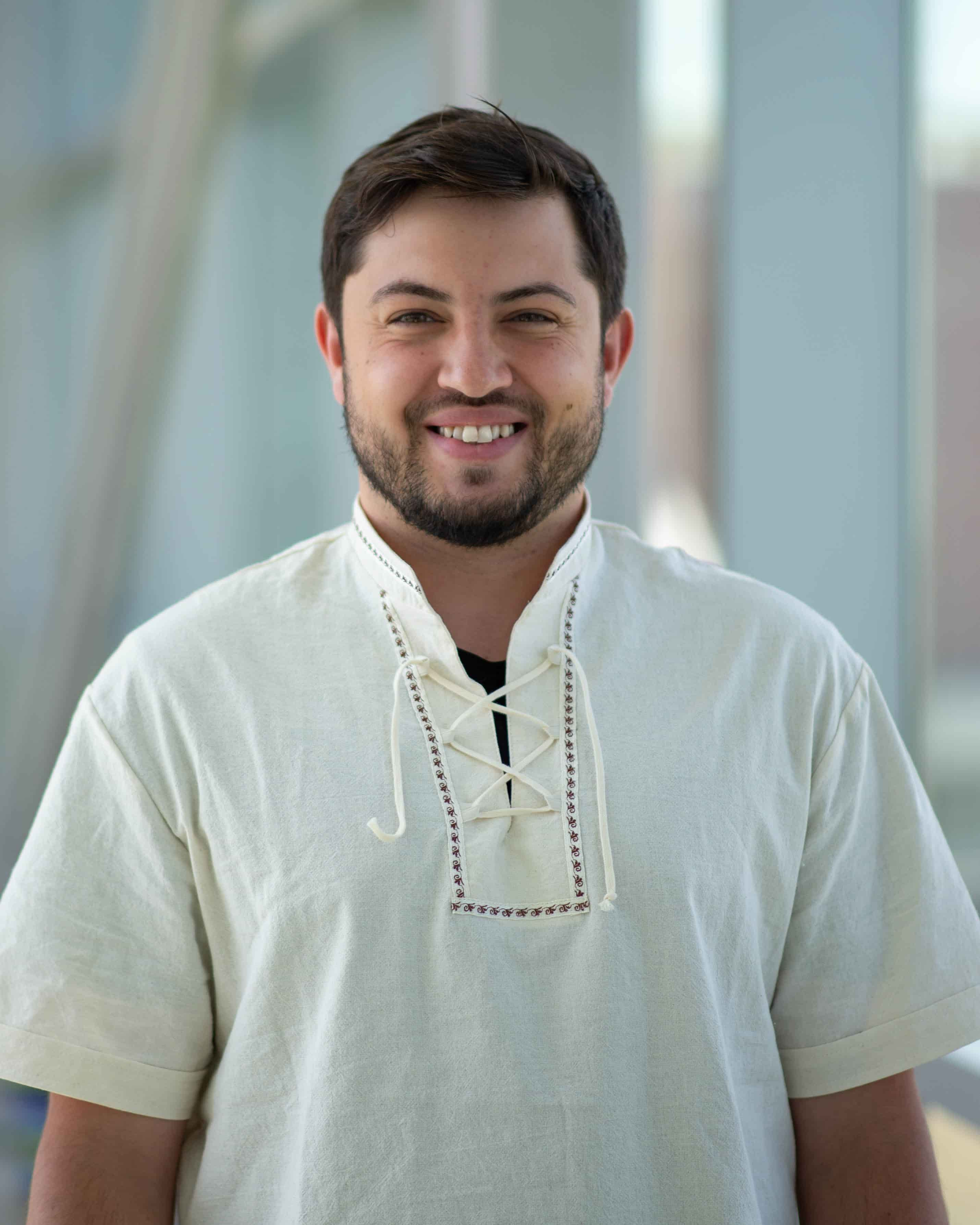
ALMC Personnel
Advanced Light Microscopy Core (ALMC)
The Advanced Light Microscopy Core (ALMC) is a user facility that serves the Anschutz Medical Campus scientific community by offering a wide variety of microscopy imaging techniques, from confocal imaging to super-resolution microscopy to whole organ
cleared tissue imaging. Training and support for all ALMC instruments is provided at no charge for academic users. The ALMC is located on the 1st floor in Research Complex 1 North (RC1-N) and on the 7th floor in Research Complex 2 (RC2).
The ALMC can assist you with fluorescent and label-free imaging of various cellular structures or tissues in several ways including, but not limited to:
- Confocal Imaging – for obtaining crisp images with good z-resolution and for 3D imaging (diffraction limited resolution: 200nm in-plane and 500nm axial).
- TPE/SHG (two-photon excitation / second harmonic generation) Imaging – for visualization of collagen/elastin and weak auto-fluorescent label-free structures to depths that can exceed hundreds of microns.
- TIRF (total internal reflection fluorescence) Imaging – for imaging and monitoring events happening on the cell membranes (<100nm depth).
- Real Time Confocal Imaging – for live cell imaging and monitoring fast dynamics within the cell, by using spinning disk confocal (up to 30fps).
- CARS (coherent anti-stokes raman spectroscopy) Imaging – for visualization of lipids, myelin and certain other label-free cellular components.
- FCS/FCCS (fluorescent (cross-)correlation spectroscopy) – for determining proteins diffusion properties, local concentration, size, binding and clustering.
- RICS/N&B (raster imaging correlation spectroscopy / number & brightness analysis) – for determining proteins diffusion properties, clustering and binding stoichiometry.
- Spectral detection imaging with un-mixing up to 8 different stains or fluorescent proteins spectra.
- FLIM (fluorescent lifetime imaging) – for imaging based on fluorophore photon emission rather than standard wavelength (“color”) imaging; for NADH/FAD and other intrinsic auto-fluorescence entities in cells or tissue
- FRET (förster resonance energy transfer) Imaging – for monitoring molecular interactions and proteins binding (FLIM-based FRET available).
- STED (stimulation emission depletion) super-resolution imaging with resolution beyond diffraction limit, down to 50nm in-plane resolution, two-channel for co-localization.
- 3D STORM/PALM (super-resolution localization) that can achieve 20nm in-plane resolution and 50nm axial resolution.
Let us see if we can help you.
To set up an account, learn how to reserve an instrument or for assistance with imaging needs for your project, please contact the ALMC at almc@cuanschutz.edu or Radu Moldovan (303-724-3649, radu.moldovan@cuanschutz.edu).
If you already have an account, you may proceed directly to the reservation page.
Rates
Our rates depend on your lab affiliations, as follows:
- NTC rate: $30/hr advanced+confocal, $20/hr non-confocal (for NeuroTechnological Center partnering departments)
- DRC rate: $30/hr advanced+confocal, $20/hr non-confocal (for Diabetes Research Center members)
- CU-AMC rate: $45/hr advanced+confocal, $25/hr non-confocal (all other on Anschutz Medical Campus)
- External Academic rate: $100/hr (non-Anschutz academic, i.e. CU Boulder)
- External rate: $100/hr (private sector, industry)
- Training (2 hours) is free for academic users
Acknowledgements
Please acknowledge the ALMC facilities in all publications and grant applications where our equipment and/or personnel have facilitated the work. If core equipment was used and data published, please note the core equipment in the materials and methods. Please include ALMC personnel as co-authors on your publications when they make a significant contribution to your research.Feel free to copy and paste the following to acknowledge the core:
| "We would like to thank the personnel from the University of Colorado Anschutz Medical Campus Advanced Light Microscopy Core for imaging assistance. Imaging was performed in the Advanced Light Microscopy Core facility of the NeuroTechnology Center at the University of Colorado Anschutz Medical Campus, which is supported in part by Rocky Mountain Neurological Disorders Core Grant (P30NS048154) and by Diabetes Research Center Grant (P30 DK116073)." |


