Instruments
 |
Imaging Techniques |
Features
Features
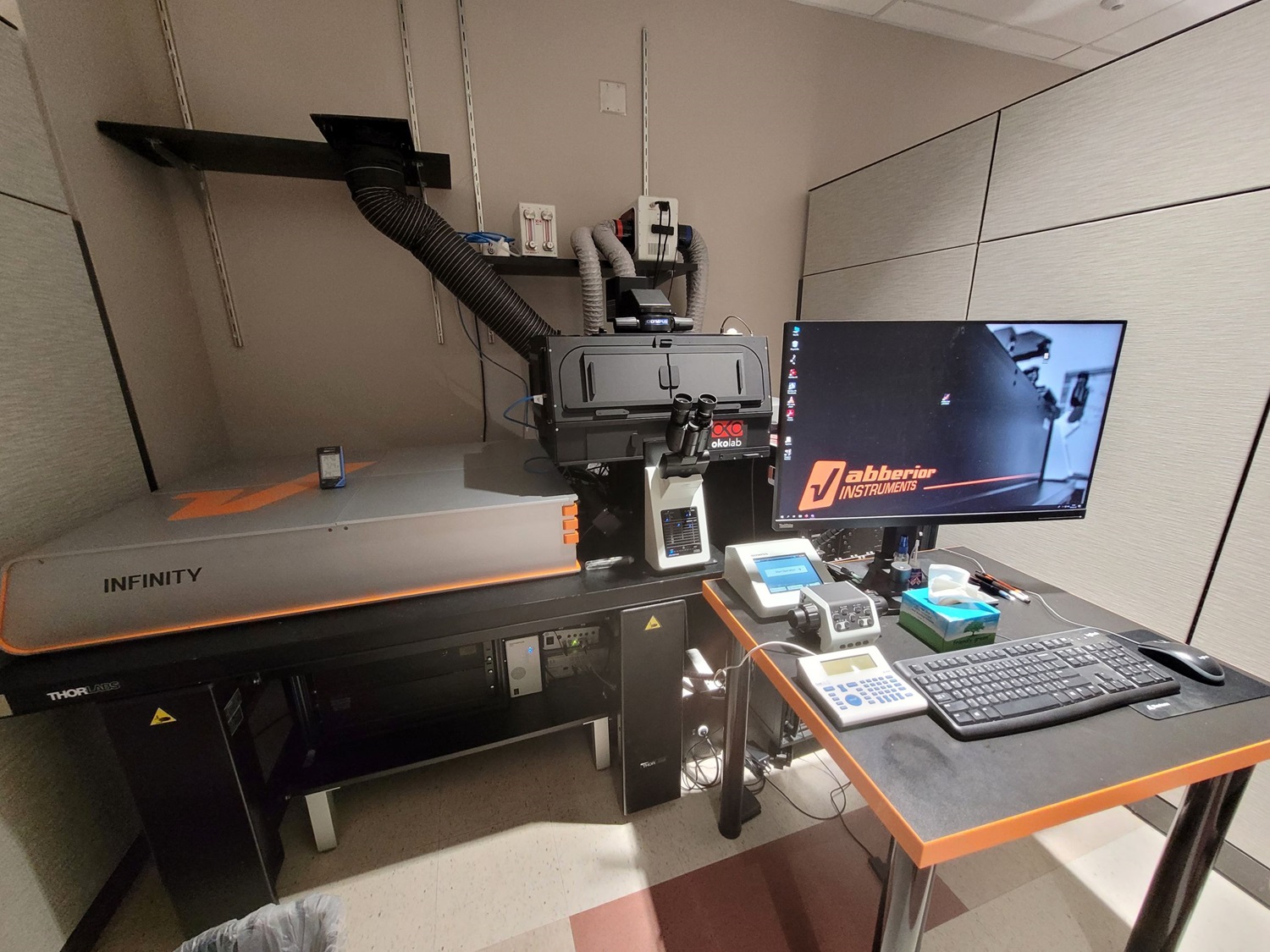
The Abberior Infinity Line is an inverted microscope that offers 2D and 3D stimulated emission depletion (STED) imaging as well as laser scanning confocal imaging. In 2D STED lateral resolutions down to 30nm (axially 600nm), and in 3D STED isotropic resolution down to 80nm can be achieved with photostable fluorophores. This microscope is equipped with adaptive optics for deep 3D STED. Adaptive illumination with RESCue or DyMIN methods reduce photobleaching. The PicoQuant MultiHarp 150 TCSPC unit allows for florescence lifetime measurements. This microscope is equipped with an environmental enclosure and a live cell stage insert. A piezo Z-axis stage allows for fast and accurate z-stacks.
Software
- Lightbox
- Imspector
- SVI Huygens Essentials
Light Sources
- STED / Laser scanning confocal:
- 405nm CW laser diode and 440nm, 485nm, 561nm, and 640nm pulsed lasers diodes
- 595nm and 775nm pulsed STED laser
- Spatial-light modulators for generating optimal 2D and 3D STED beam profiles
- Epi-fluorescence (eye pieces):
- CoolLED pE-4000
Detection
- 4 freely tunable channels (RAINBOW detection unit) from 415nm to 750nm, 3 with avalanche photo diodes (APDs) and one with a multi-element MATRIX detector
- Picoquant MultiHarp150 for FLIM imaging in 4 channels, and TIMEBOW module for FLIM based channel separation and visualization.
- Deformable mirror for spherical aberration correction when imaging volumetric samples
- Multi element MATRIX detector to improve signal-to-background ratio.
Objectives
- 20x / NA 0.80, Air
- 60x / NA 1.30, Silicone Oil
- 60x / NA 1.42, Oil
Location: 1st floor RC1N, room P18-1304
 | Imaging Techniques |
Features
Features
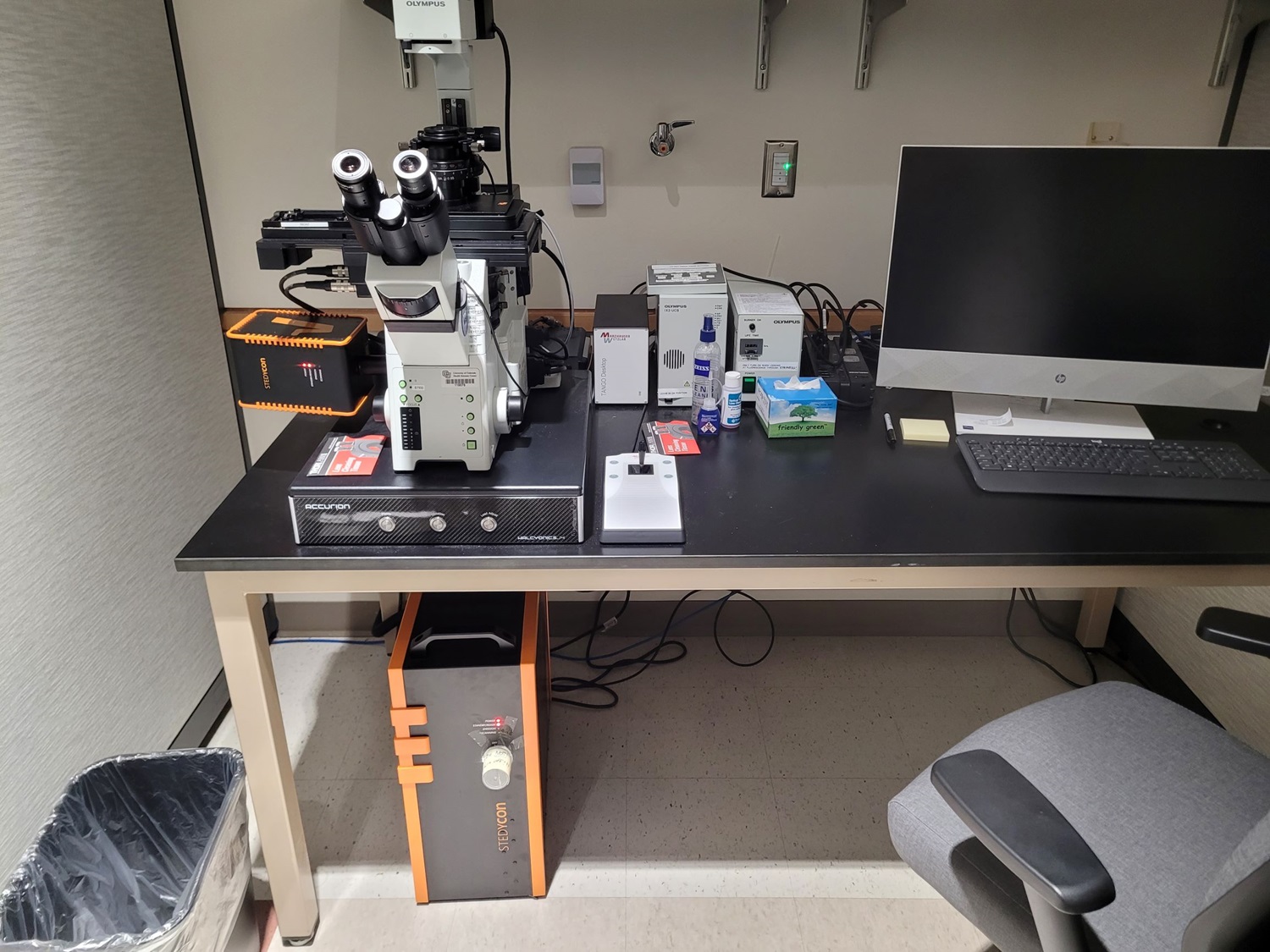
The Abberior STEDYCON is a user-friendly microscope that allows for 2D stimulated emission depletion (STED) super-resolution microscopy in up to 3 imaging channels with a lateral resolution down to 40nm (axially 600nm). The STEDYCON also offers laser scanning confocal imaging in up to 4 channels. A piezo Z-axis stage allows for fast and accurate z-stacks.
Software
- STEDYCON web interface
- SVI Huygens Essentials
Light Sources
- STED / Laser scanning confocal:
- 405nm CW laser diode and 485nm, 561nm, and 640nm pulsed lasers diodes
- 775nm pulsed STED laser
- Epi-fluorescence (eye pieces):
- Excelitas X-Cyte Xylis LED
Detection
- Laser Scanning Confocal:
- 4 avalanche photodiode (APD) channels in single photon counting mode with fixed spectral bandpass filters
- blue (420nm – 480nm), green (505nm-545nm), red (575nm – 625) and far-red (650nm – 700nm)
- 4 avalanche photodiode (APD) channels in single photon counting mode with fixed spectral bandpass filters
- STED:
- Up to 3 STED channels using 775nm STED laser and far-red, red and long-Stokes-shift (excitation with 485nm, fluorescence in the red or far-red channel) fluorophores. STED and confocal channels can be imaged in the same acquisition.
Objectives
- 10x / NA 0.4, Oil
- 20x / NA 0.85, Oil
- 40x / NA 1.35, Oil
- 100x / NA 1.45, Oil
Location: 1st floor RC1N, room P18-1304
 |
Imaging Techniques |
Features
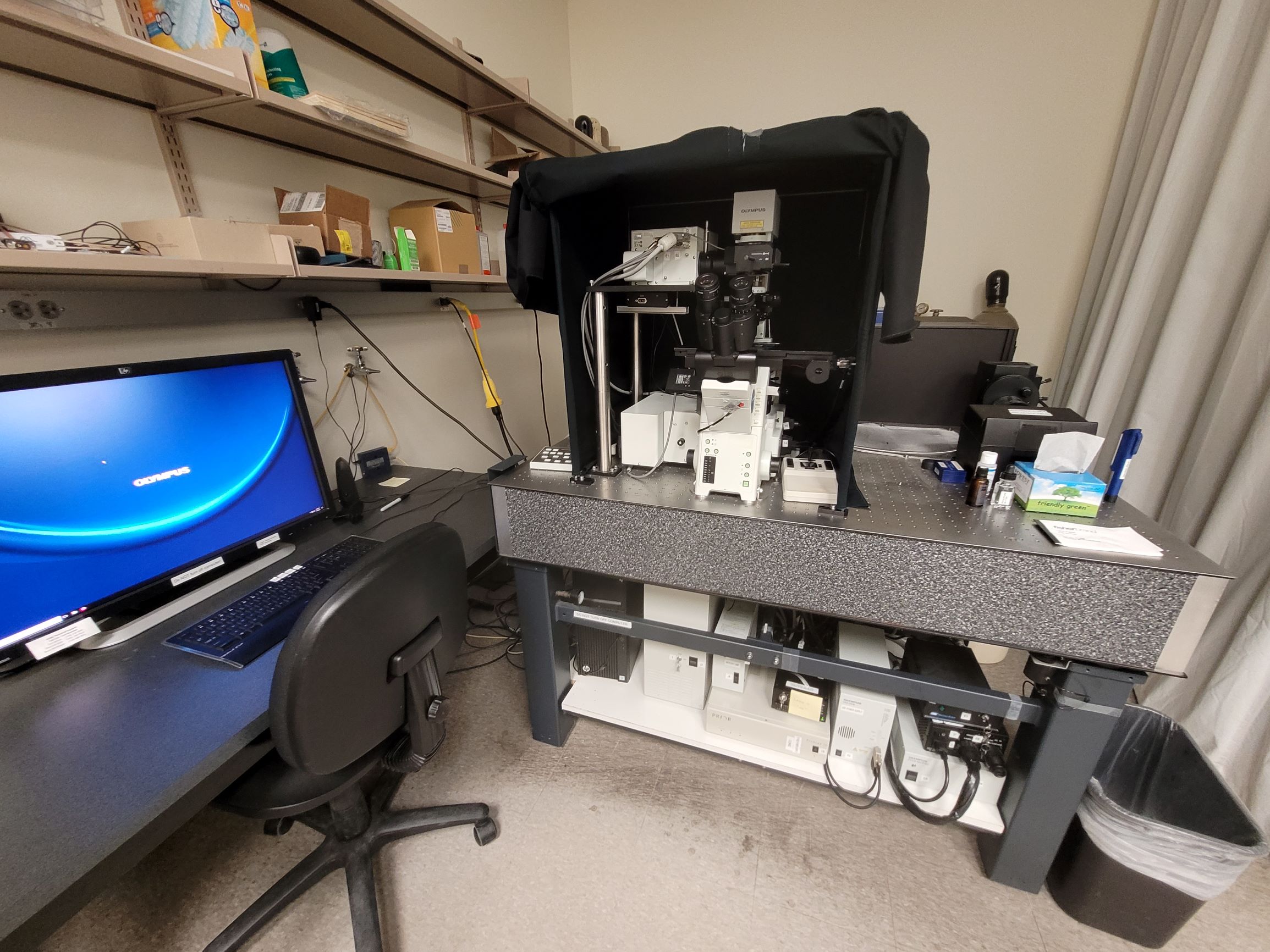
The Olympus FV1000 is an inverted laser scanning confocal microscope equipped with a live cell stage insert. Spectral FRET imaging and analysis is available. The APE picoEmerald laser system allows for coherent anti-Stokes Raman scattering (CARS) imaging.
Acquisition Software
- FV10-ASW 4.2
- APE picoEmerald 4.10 (CARS)
Light Sources
- Laser scanning confocal:
- 405nm, 458nm, 488nm, 515nm, 543nm, and 635nm CW lasers
- CARS:
- APE picoEmerald, pulsed picosecond optical parametric oscillator with tuning range from 700nm–960nm, and a fixed 1031nm laser
- Epi-fluorescence (eye pieces):
- Mercury Halide Lamp
Detection
- Laser Scanning Confocal:
- 3 PMT channels with filter based spectral selection to image up to 3 confocal channels simultaneously in the range from 430nm – 750nm (unlimited channels in sequential mode)
- CARS:
- 2 non-descanned PMT channels in forward direction:
- bandpass filters from 2500 cm-1 - 2950 cm-1 or 1775 cm-1 - 2240 cm-1 with autofluorescence
- 2 non-descanned PMT channels in epi- direction:
- bandpass filter for CARS imaging from 2500 cm-1 to 2950 cm-1
- 2 non-descanned PMT channels in forward direction:
- Brightfield/DIC imaging:
- 1 transmitted light PMT channel
Objectives
- 4x / NA 0.13, Air
- 10x / NA 0.40, Air
- 20x / NA 0.75, Air
- 30x / NA 0.10, Silicone
- 60x / NA 1.20, Water
- 100x / NA 1.40, Oil
Location: 7th floor RC2, room P15-7251
Note: RESTRICTED USE for CARS imaging, only with ALMC personnel assistance!
 |
Imaging Techniques |
Features
Features
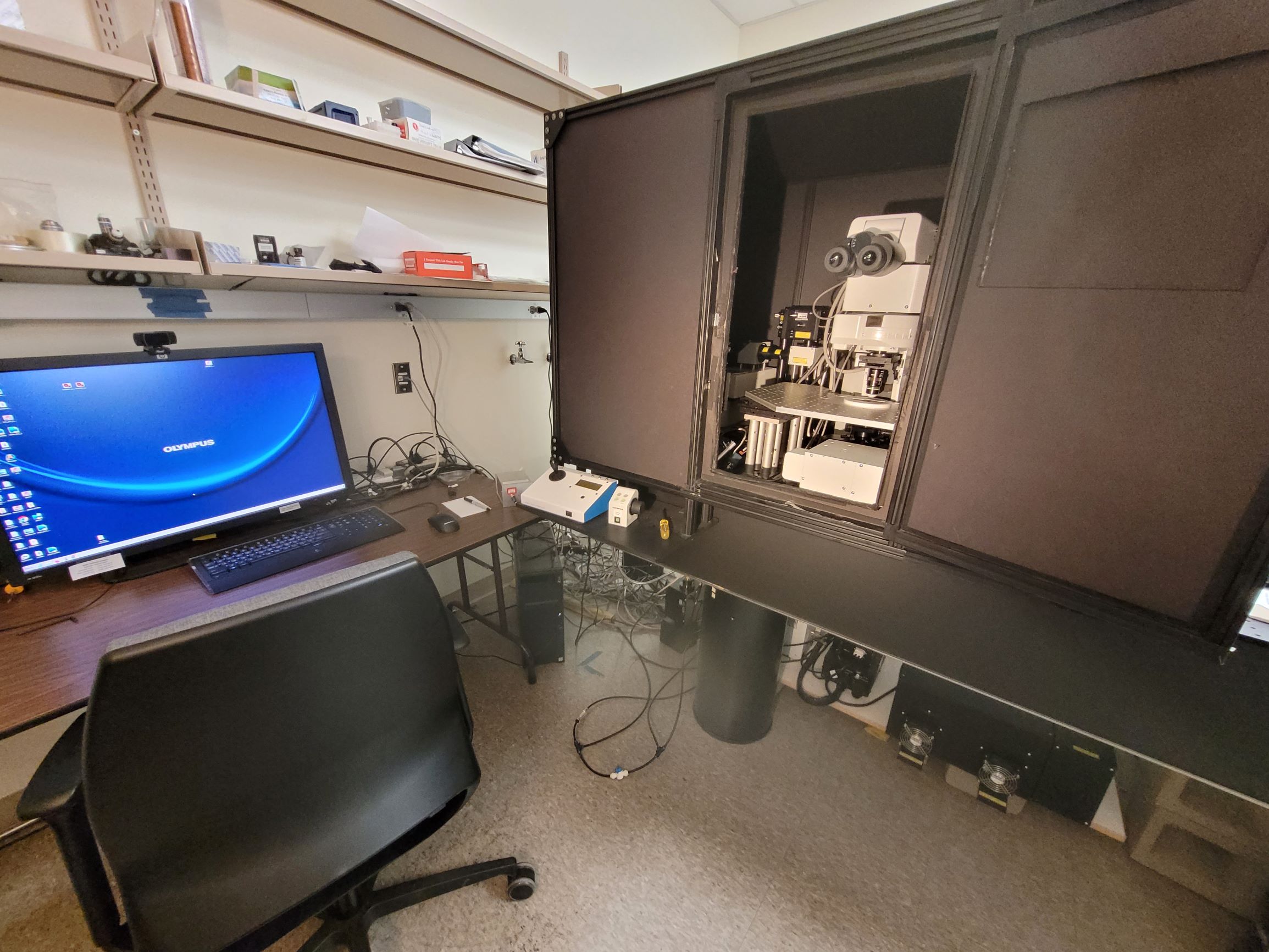
The Olympus FV1000 is an upright microscope that offers laser scanning confocal and multiphoton microscopy. It has an optional large area PMT DIVER detector for efficient detection in forward direction of second harmonic generation (SHG), two photon excitation (TPE), fluorescent lifetime imaging microscopy (FLIM) or fluorescence in deep tissue imaging experiments. The microscope is housed within a light-tight enclosure and has two custom stages that accommodate a large variety of samples, from normal microscope slides to in-vivo experiments.
Software
- FV10-ASW 4.2
- ISS VistaVision (FLIM)
Light Sources
- Laser scanning confocal:
- 405nm, 458nm, 488nm, 515nm, 543nm, and 635nm CW lasers
- SHG, 2p fluorescence:
- SpectraPhysics Insight X3, pulsed femtosecond laser with a tuning range from 680nm-1300nm, and a fixed laser line at 1045nm
- Epi-fluorescence (eye pieces):
- Mercury Halide Lamp
Detection
- Laser Scanning Confocal:
- 3 PMT channels with filter based spectral selection to image up to 3 confocal channels simultaneously in the range from 430nm – 750nm (unlimited channels in sequential mode)
- SHG and 2p fluorescence:
- 4 non-descanned PMT channels in the epi-direction with the following filter cubes available for fluorescence:
- blue (420nm – 460nm), green (495nm – 540nm), red (575nm-630nm), and far-red (650nm – 700nm) channels
- NADH (430nm – 490nm) and FAD (515nm – 595nm)
- SHG (370nm – 410nm) and two-photon autofluorescence (450nm – 650nm)
- 2 non-descanned PMT channels in forward direction for SHG imaging at 800nm or 1045nm
- Single shot HDR imaging (imaging same fluorescence channel with PMTs to combine two native 12bit channels to one 16bit imaging channel)
- 4 non-descanned PMT channels in the epi-direction with the following filter cubes available for fluorescence:
- Brightfield/DIC imaging:
- 1 transmitted light PMT channel
- DIVER detector for SHG, THG, and FLIM (with ISS FastFLIM unit)
- 1 transmitted light PMT channel
Objectives
- 10x / NA 0.30, Water
- 10x / NA 0.50, Glycerol (refractive index range: 1.33 – 1.515)
- 16x / NA 0.80, Water
- 20x / NA 1.00, Water
- 40x / NA 0.80, Water
- 60x / NA 1.20, Water
Location: 7th floor RC2, room P15-7251
 | Imaging Techniques |
Features
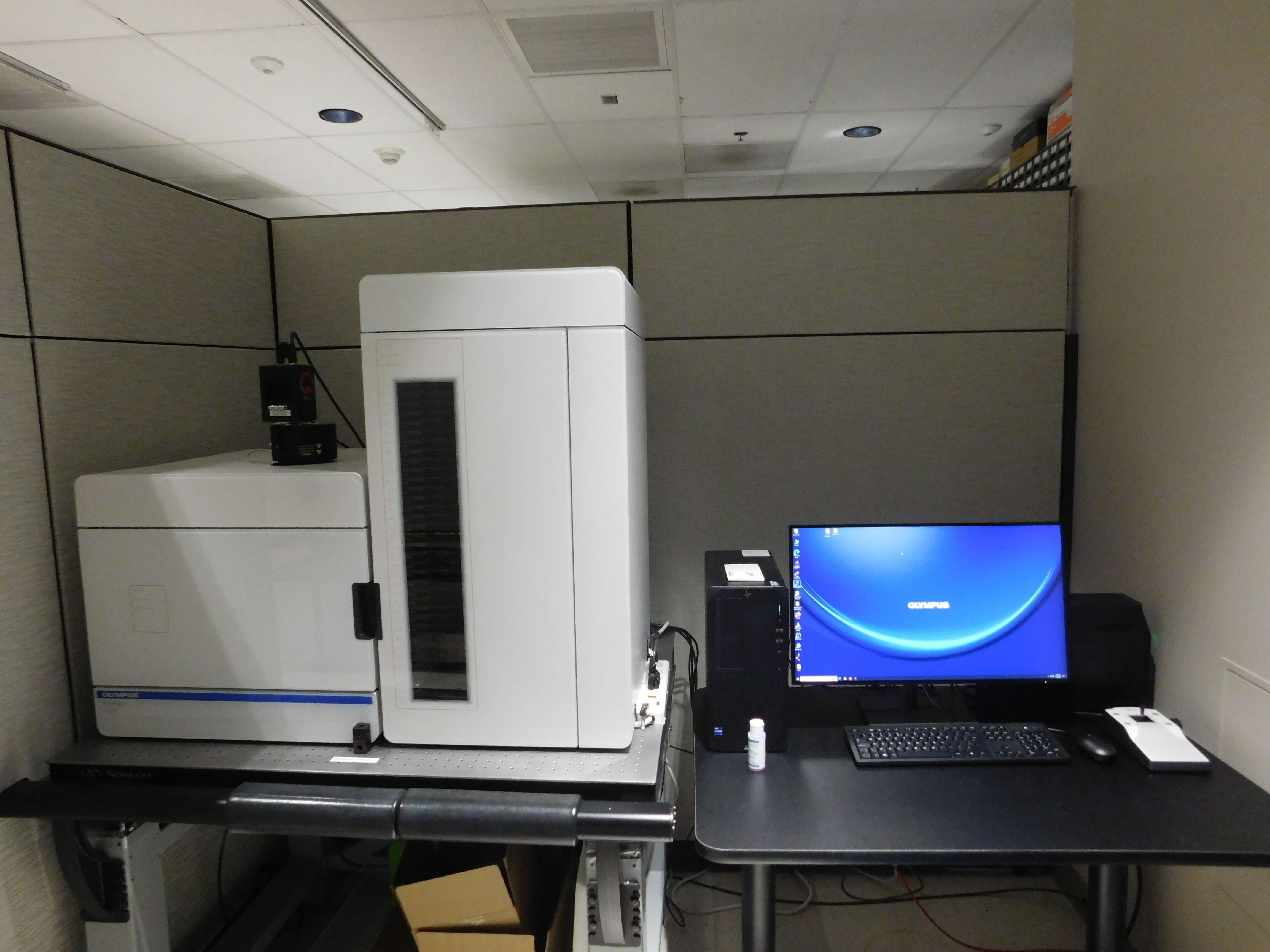
The Olympus VS200 is a high throughput slide scanning microscope made for imaging large fields of view or entire slides. It can image in brightfield, fluorescence, darkfield, and phase contrast modes. This microscope has an automated slide loader system that can hold 210 slides. An automated oil dispensing system allows imaging with immersion objectives.
Acquisition Software
- Olympus OlyVIA V4.1
Light Sources
- Epi-fluorescence:
- Excelitas X-Cite II XT730L White Light LED
- Penta-band excitation filters for Dapi (352nm – 404nm), FITC (460nm – 487nm), TRITC (541nm – 568nm), CY5 (624nm – 647nm), CY7 (719nm – 751nm) channels
- SpectraSplit 7 bandpass excitation filters for Opal dyes
- Brightfield:
- Olympus True Color LED
Detection
- Epi-fluorescence:
- Hamamatsu Orca-Fusion Gen III sCMOS
- Penta-band emission filters for Dapi (413nm – 451nm), FITC (497nm – 532nm), TRITC (577nm – 613nm), CY5 (657nm – 704nm), CY7 (766nm – 855nm) channels
- SpectraSplit 7 band pass emission filters for Opal dyes
- Brightfield:
- Integrated 2/3” CMOS (3.45 μm pixel size)
Objectives
- 2x / NA 0.06, Air
- 4x / NA 0.16, Air
- 10x / NA 0.40, Air
- 20x / NA 0.80, Air
- 60x / NA 1.42, Oil
Location: 1st floor RC1N, room P18-1303
 |
Imaging Techniques |
Features
Features
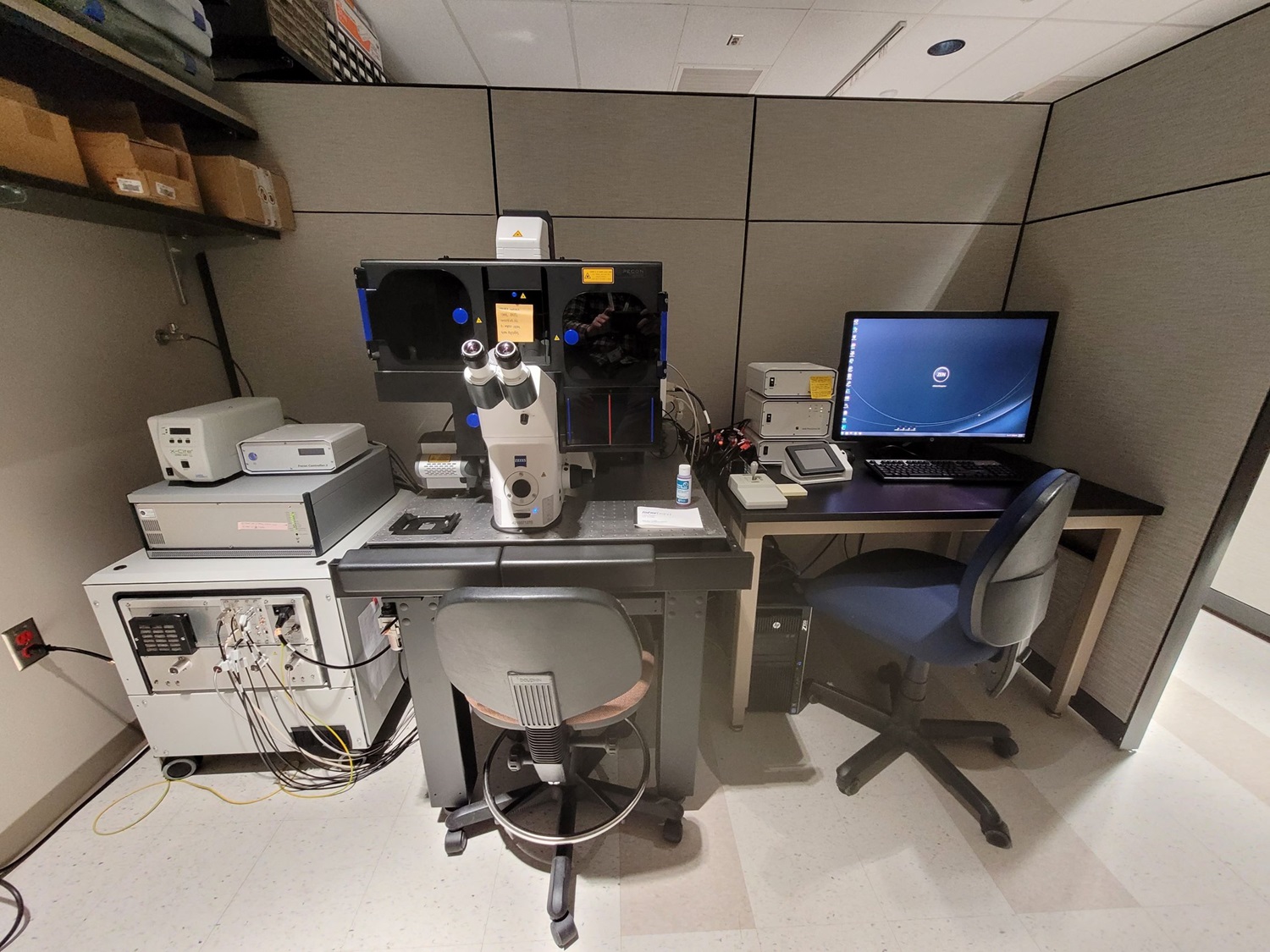
The Zeiss Elyra P.1 is a widefield microscope that is designed for PALM/STORM super resolution imaging, with axial resolution down to 30nm. It offers Epi, HiLo and TIRF illumination modes. It has a focus stabilization system to be used during long PALM/STORM or TIRF live cell image acquisitions.
Software
- Zen Black
Light Sources
- Epi/HiLo/TIRF illumination:
- 405nm, 488nm, 561nm CW lasers
- Epi-fluorescence (eye pieces):
- Excelitas X-Cite 120PC Q Mercury Vapor Lamp
Detection
- Andor iXon+ 897 EMCCD camera and the following filter cubes:
- blue (420nm – 480nm), green (495nm – 550nm), red (570nm - 620nm) and far-red (655nm – 720nm)
Objectives
- 10x / NA 0.30, Air
- 63x / NA 1.40, Oil
- 100x / NA 1.46, Oil
Location: 1st floor RC1N, room P18-1303
 | Imaging Techniques |
Features
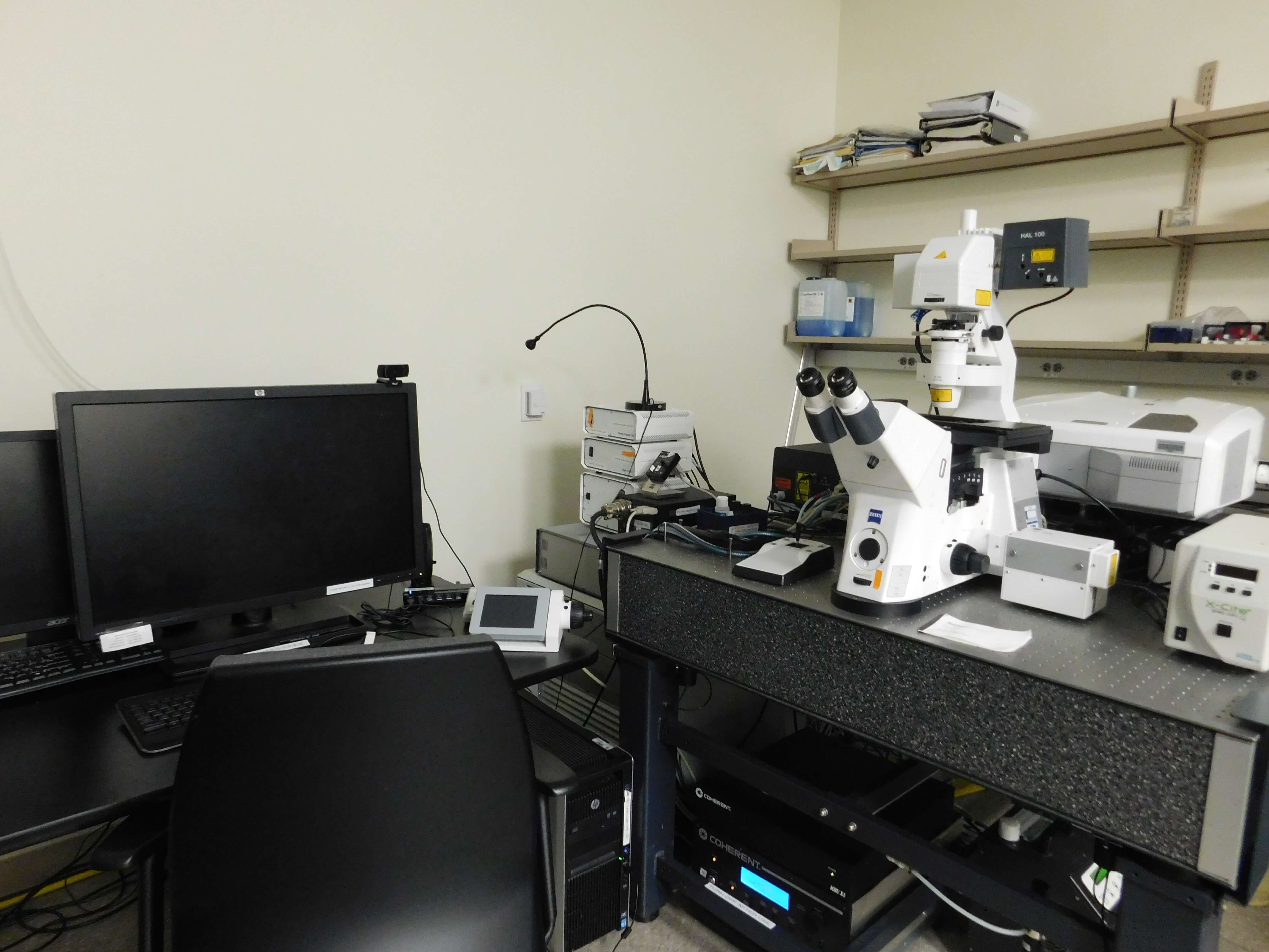
The Zeiss LSM780 is an inverted laser scanning confocal microscope capable of hyperspectral imaging that supports multicolor fluorescent samples. The 32-Channel GaAsP QUASAR detector allows for tunable spectral detection. This microscope is equipped with a live cell stage insert and a focus stabilization system. Fluorescence correlation spectroscopy (FCS) and raster image scanning correlation spectroscopy are supported. The tunable femtosecond laser allows second harmonic generation (SHG), two photon excitation (TPE), fluorescence lifetime imaging (FLIM) and FLIM based Förster resonant energy transfer (FRET) imaging.
Acquisition Software
- Zen Black
- ISS Vista Vision (FLIM)
Light Sources
- Laser scanning confocal:
- 458nm, 488nm, 514nm, 561nm, and 633nm CW lasers
- SHG, 2p Fluorescence, FLIM:
- Coherent Chameleon Ultra 2, pulsed femtosecond laser with tuning range from 680nm–1080nm
- Epi-fluorescence (eye pieces):
- Excelitas X-Cite 120PC Q Mercury Vapor Lamp
Detection
- Laser Scanning Confocal/2p Fluorescence/FCS:
- Freely tunable imaging channels from 371nm – 759nm for imaging in up to 3 confocal channels simultaneously (unlimited channels in sequential mode)
- Hyperspectral imaging with QUASAR detection unit with 32 channels
in 410nm – 696nm range (possible binwidths: 3nm, 4.4nm,
8.9nm,17.8nm, 26.7nm, 35.5nm, GaAsP detectors), plus 2 PMT detectors in the UV and far-red to extend the spectrum
- SHG/2p Fluorescence:
- 2 non-descanned PMTs
- Filter cubes available for the following channel combinations
(NADH/FAD, SHG/TPE, Nile Red, TRITC) - Laser scanning confocal channels are utilizable in same acquisition
- FLIM:
- 2 descanned single photon counting PMTs for imaging with an ISS FastFLIM unit or a Becker&Hickl TCSPC card
- Brightfield/DIC imaging:
- 1 transmitted light PMT channel
Objectives
- 10x / NA 0.30, Air
- 20x / NA 0.80, Air
- 40x / NA 1.20, Water
- 63x / NA 1.40, Oil
- 100x / NA 1.40, Oil
Location: 7th floor RC2, room P15-7251
 | Imaging Techniques |
Features
The Zeiss LSM980 is an inverted laser scanning confocal and two photon microscope. It is equipped with a 32-Channel GaAsP QUASAR detector for tunable spectral detection, spectral FCS, and hyperspectral imaging. It also features an Airyscan 2 detector for super resolution imaging, multi-line scanning, and spatial FCS. The tunable femtosecond laser allows for SHG, TEP, FLIM, and FLIM based FRET imaging. Two GaAsP BiG.2 non-descanned detectors are available for sensitive SHG/TPE imaging. The CMOS camera allows large area overviews with epi-fluorescence in the DAPI/FITC/TRITC channels. Paired with the AI Sample Finder, this option offers efficient imaging workflows. The microscope is equipped with a live cell stage insert and a focus stabilization system.
Acquisition Software
- Zen Blue 3.10
Light Sources
- Laser scanning confocal:
- 405nm, 445nm, 488nm, 514nm, 561nm, and 639nm CW lasers
- SHG/TPE/FLIM:
- SpectraPhysics Mai Tai, pulsed femtosecond laser with tuning range from 690nm–1040nm
- Epi-fluorescence:
- Excelitas X-Cite XYLIS LED with excitation from 380nm - 770nm
Detection
- Laser Scanning Confocal/TPE/FCS:
- Freely tunable imaging channels from 371nm – 759nm for imaging in up to 3 confocal channels simultaneously (unlimited channels in sequential mode)
- Hyperspectral imaging with QUASAR detection unit with 32 channels in 410nm – 696nm range (possible binwidths: 3nm, 4.4nm, 8.9nm,17.8nm, 26.7nm, 35.5nm, GaAsP detectors), plus 2 PMT detectors in the UV and far-red to extend the spectrum
- Super-resolution imaging with Airyscan 2 detection unit with 8Y Multiplexing for 2x or 8x parallelization. Spatially resolved FCS is possible with the Dynamics Profiler software package
- SHG/2p Fluorescence:
- 2 non-descanned BiG.2 GaAsP detectors with filter cubes for NADH/FAD, SHG/TPE, FITC/TRITC
- Airyscan 2 can be used with 2p fluorescence for super-resolution imaging
- Laser scanning confocal channels are utilizable in same acquisition
- FLIM:
- 2 descanned single photon counting PMA Hybrid 40 mod detectors with a MultiHarp 150 TCSPC card from PicoQuant. Channels: 464nm - 500nm and 524nm - 576nm
- Brightfield/Epi-fluorescence:
- 1 transmitted light PMT channel
- Axiocam 305 mono R2
Objectives
- 2.5x / NA 0.085, Air
- 10x / NA 0.30, Air
- 20x / NA 0.80, Air
- 40x / NA 1.20, Water
- 63x / NA 1.40, Oil
Location: 7th floor RC2, room P15-7244
Acknowledgements
"We would like to thank the personnel from the University of Colorado Anschutz Medical Campus Advanced Light Microscopy Core for imaging assistance. Imaging was performed in the Advanced Light Microscopy Core facility of the NeuroTechnology Center at the University of Colorado Anschutz Medical Campus, which is supported in part by Rocky Mountain Neurological Disorders Core Grant (P30NS048154) and by Diabetes Research Center Grant (P30 DK116073). Usage of the Zeiss LSM 980 was supported by S10 OD036220."
 |
Imaging Techniques |
Features
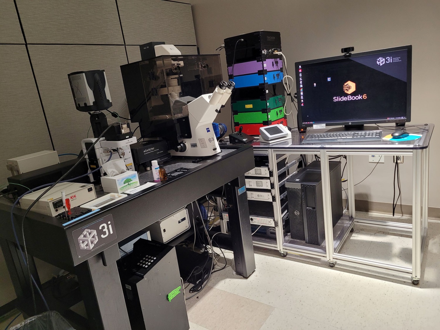
The 3I MARIANAS is an inverted spinning disk confocal microscope fit for long term and fast live cell imaging. This microscope is equipped with an environmental enclosure and live cell stage insert, including a focus stabilization system. A vector scanner is available to use for photomanipulation experiments. A separate camera is included for live cell FURA2 ratiometric calcium imaging. A piezo Z-axis stage allows for fast and accurate z-stacks.
Software
- Slidebook 6
Light Sources
- Spinning disk confocal:
- 405nm, 445nm, 488nm, 515nm, 561nm, and 640nm CW lasers
- 3i mSwitcher and Vector for photomanipulation with any laser line
- Epi-fluorescence:
- Sutter Lambda DG4 lamp
Detection
- Spinning disk confocal:
- Yokogawa CSU-X1 spinning disk unit with Andor iXon 888 EMCCD
- Imaging channels: Dapi (422nm to 467nm), Fitc (500nm to 550nm), Tritc(580nm to 653nm), Alexa647 (677nm to 722nm), CFP (465nm to 500nm), YFP (529nm to 556nm)
- Epi-fluorescence:
- Photometrics CoolSNAP HQ2 CCD
- Imaging channels: CFP, GFP, RFP, Fura2
Objectives
- 10x / NA 0.30, Air
- 20x / NA 0.75, Air
- 40x / NA 0.75, Air
- 40x / NA 1.30, Oil
- 63x / NA 1.40, Oil
- 100x / NA 1.40, Oil
Location: 1st floor RC1N, room P18-1304
 | Imaging Techniques |
Features
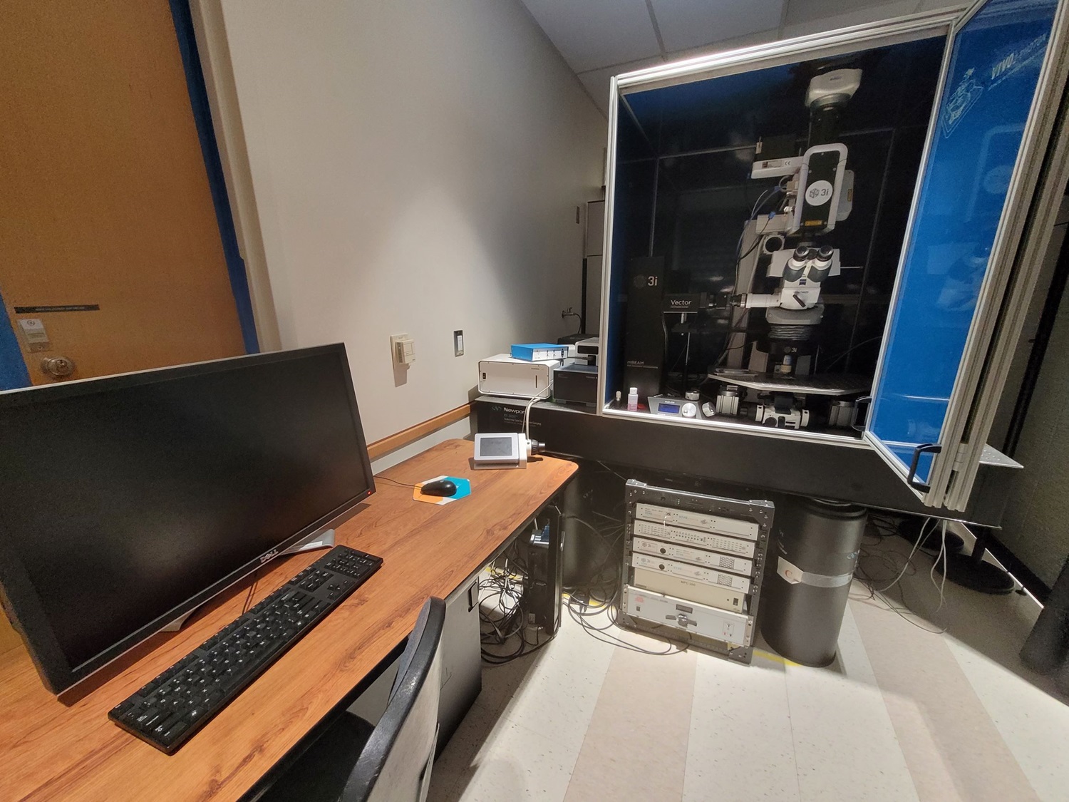
The 3I VIVO is an upright microscope that offers spinning disk confocal. A large stage accommodates long-term imaging for in/ex-vivo.
Software
- Slidebook 6
Light Sources
- Spinning disk confocal:
- 405nm, 488nm, 561nm CW lasers
- Epi-fluorescence (eye pieces):
- Excelitas X-Cite 120 Q Mercury Vapor Lamp
Detection
- Spinning disk confocal:
- Yokogawa CSU-X1 spinning disk unit with Photometrics Evolve 16-bit EMCCD
- Imaging channels: Dapi (422nm to 467nm), Fitc (500nm to 550nm), Tritc(580nm to 653nm)
Objectives
- 20x / NA 1.0, Water
- 20x / NA 1.0, refractive index 1.38 (ScaleA2 Tissue Clearing)
- 63x / NA 0.90, Water
Location: 1st floor RC1N, room P18-1304
 |
Features
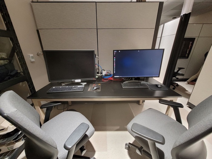
Two computers with a variety of image analysis software are available to all users!
Available software packages
Imaris 9
- Vantage
- Measurment Pro
- Coloc
Imaris 9 & 10
- Vantage
- Measurement Pro
- Coloc
- Cell
- Filament Tracer
- Batch
SVI Huygens
- Deconvolution
- Confocal, STED, STED 3D
- Restoration
- Chromatic Aberration, Crosstalk, Object Stabilizer, Tile Stitching
- Visualization
- Twin Slicer, Ortho Slicer
- Colocalization
- Abberior Bundle
Open Source and Free Software
- FIJI
- QuPath
- NIS-Elements
- OIViewer (Available Locally)
- Olympus: OlyVIA, FV31S-SW, FV10-ASW 4.2
- FLIM analysis: VistaVision 4.2 (Available Locally), Leica Falcon Software (Available Locally), FLIMFit 5.0.3
- Slidebook 6
- Matlab
- Zeiss: ZEN, Arivis (Available Locally)
Location: 1st floor RC1N, room P18-1304
The ALMC houses a wide range of imaging systems, each with unique advantages. Please contact us so we can help find the best imaging solution for your research.
CMS Login