Skip to content
University of Colorado Anschutz Medical Campus CU Anschutz
  • Quick Links
 

Tools & Resources

  • Events Calendar
  • Newsroom
  • Strauss Health Sciences Library
  • Department A-Z Directory
  • Campus Directory
  • Leadership
  • Faculty & Staff Resources
  • Supporter & Alumni Resources
  • Student Resources
  • Mental Health Resources
  • Campus Map
  • University Policies
  • Give Now

CU Campuses

  • CU Anschutz Medical Campus
  • CU Boulder
  • CU Colorado Springs
  • CU Denver
  • CU System
  • CU Online

CU Anschutz Medical Campus

  • School of Dental Medicine
  • Graduate School
  • School of Medicine
  • College of Nursing
  • Skaggs School of Pharmacy and Pharmaceutical Sciences
  • Colorado School of Public Health
  • Home
  • My Practice
    • Dr Eric McCarty
    • My Team
    • Practice Locations
  • Practice Expertise
    • Knee
      • ACL Injuries
      • Meniscal Tears
      • Articular Cartilage Injuries
      • Medial Collateral Ligament Injuries
      • Posterior Cruciate Ligament Injuries
      • Lateral Collateral Ligament Injuries
      • Patella Tendon Rupture
      • Quadricep Tenon Rupture
    • Shoulder
      • Rotator Cuff Tear
      • Labral Tear
      • Shoulder Instability
      • Shoulder Separation
      • Biceps Tendonitis
      • Acromion Clavicular Joint
      • Sternoclavicular Joint
      • Glenohumeral Osteoarthritis
      • Clavicle Fracture
    • Elbow
      • Distal Biceps Rupture
      • Medial Epicondylitis
      • Lateral Epicondylitis
      • Elbow Loose Bodies
    • Trauma
      • Distal Biceps Rupture
      • Quadricep Tendon Rupture
      • Patella Tendon Rupture
      • Achilles Tendon Rupture
      • Shoulder Separation
      • Pectoralis Rupture
      • Triceps Rupture
      • Clavicle Fracture
      • Fractures
  • Patient Information
    • Preparing for Surgery
    • Home Exercise Videos
    • Post Operative Information
    • Rehabilitation Protocols
    • Patient Reviews
  • Educational Resources
    • Patient Education Videos
    • Research
  • Media
    • Videos
    • In the News
    • Awards
  • Contact Dr. McCarty
    • How to Schedule
    • Contact the Team
    • Practice Locations
University Quick Links
Eric McCarty, MD
Orthpaedic Surgeon
Sports Medicine
Eric McCarty, MD,  CU Sports Medicine
Book an Appointment
  • Denver (303) 694-3333
  • Boulder (303) 315-9900
  • TRAUMA

    Acute Injury Pain

    Trauma Conditions

    Distal Biceps Rupture
    Quadricep Tendon Rupture
    Patella Tendon Rupture
    Tricep Rupture
    Pectoralis Rupture
    Achilles Tendon Rupture
    Shoulder Separation
    Clavicle Fracture
    Fractures

    Foot and Ankle Anatomy

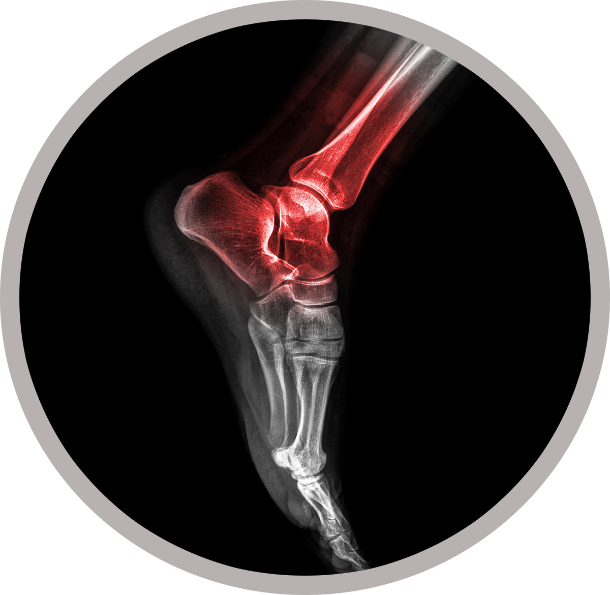
    The foot and ankle is a complex joint involved in movement and providing stability and balance to the body. The foot and ankle consists of 26 bones, 33 joints, and many muscles, tendons and ligaments.

    Bones of the Ankle

    The ankle joint connects the leg with the foot, and is composed of three bones: tibia, fibula and talus. The tibia or shin bone and fibula or calf bone are bones of the lower leg which articulate with the talus or ankle bone, enabling up and down movement of the foot.

    Three bony bumps present on the ends of the tibia and fibula form parts of the ankle joint:

    • The Medial malleolus, formed by the tibia, is found on the inside of the ankle
    • Posterior malleolus, also formed by the tibia, is found at the back of the ankle
    • Lateral malleolus, formed by the fibula, is found on the outer aspect of the ankle

    Bones of the Feet

    The foot acts as a single functional unit, but can be divided into three parts: the hindfoot, midfoot and forefoot.

    The hindfoot forms the ankle and heel and is made up of the talus bone and calcaneus or heel bone. The heel bone is the largest bone in the foot.

    The midfoot connects the hindfoot to the forefoot, and consists of one navicular bone, one cuboid bone, and three cuneiform bones. The navicular bone is found in front of the heel bone, and the cuneiform and cuboid bones are arranged in front of the navicular bone.

    These bones are connected to five metatarsal bones of the forefoot, which form the arch of the foot for shock absorption while walking or running. The forefoot is also made up of the toes or digits, formed by phalanges, three in each toe, except the big toe, which has only two phalanges. The big two has two additional tiny round sesamoid bones in the ball of the foot, which help in upward and downward movement of the toe.

    Ankle and Foot Joints

    There are 33 joints in the ankle and foot. They include:

    • Hinge joints in the ankle, which allow flexion (bending) and extension
    • Gliding joints found in the hindfoot, which allow gliding movements
    • Condyloid joints found in the forefoot and toes, which allow the flexion (bending) and extension, adduction and abduction (sideward movement)

    The joints of the foot and ankle provide stability and support the weight of the body, helping you to walk or run, and to adapt to uneven ground.

    The joint surface of all bones of the ankle and foot are lined by a thin, tough, flexible, and slippery surface called articular cartilage, which acts as a shock absorber and cushion to reduce friction between the bones. The cartilage is lubricated by synovial fluid, which further enables smooth movement of the bones.

    Soft Tissues of the Ankle and Foot

    Our feet and ankle bones are held in place and supported by various soft tissues such as cartilage, ligaments, muscles, tendons and bursae.

    Cartilage is the flexible, shiny, smooth tissue on the ends of bones that meet to form a joint. Cartilage provides cushioning between the bones allowing smooth movement.

    Ligaments are tough rope-like tissue that connect bones to other bones, and holds them in place providing stability to the joints. The Plantar fascia is the largest ligament in the foot, originating from the heel bone to the forefoot, it extends along the bottom surface of the foot and is involved in maintaining the arch of the foot. The plantar fascia ligament stretches and contracts to provide balance and strength to the foot. Lateral ligaments on the outside of the foot and medial ligaments on the inside of the foot provide stability and allow up and down movement of the foot.

    The foot is made up of 20 muscles, which help in movement. The main muscles include:

    • Anterior tibial muscle: allows up and down movement of the foot.
    • Posterior tibial muscle: supports the arch.
    • Peroneal tibial muscle: controls movement on the outside of the ankle.
    • Extensors: enable the ankle to raise the toes just before stepping forward.
    • Flexors: stabilize the toes against the floor.
    • Smaller muscles are also present to help the toes lift up and curl.
    • Tendons are soft tissues that connect muscles to bones. The largest and strongest tendon in the foot is the Achilles tendon, present at the back of the lower leg around the heel bone. Other tendons include peroneals and anterior and posterior tibialis.

    Bursae

    Bursae are small fluid filled sacs that decrease friction between tendons and bone or skin. Bursae contain special cells called synovial cells that secrete a lubricating fluid.

    The ankle and foot have a complex anatomy which functions together to help us run and walk. Any condition or injury that disrupts the way these tissues work greatly impacts the normal functioning of the foot and ankle.

    PRACTICE EXPERTISE

     Knee Injuries KNEE


    Shoulder Pain, Sports Medicine SHOULDER


    Elbow Pain, Sports Medicine ELBOW


    Acute Injuries, sports Medicine TRAUMA

    Eric C. McCarty, Orthopaedic Surgeon, Sports Medicine

    View Profile


    PATIENT EDUCATION

    Patient Education Videos

    PATIENT RESOURCES

     Your First Visit YOUR FIRST VISIT


    Practice Expertise PRACTICE EXPERTISE


    RehabProtocol  PROTOCOLS


    Surgery Information SURGERY INFORMATION


    Surgery Information IN THE NEWS

    • CU Buffs
    • Colorado Avalanche
    • CU Sports Medicine
    • CU Buffs
    • Colorado Avalanche
    • CU Sports Medicine
    • CU Buffs
    • Colorado Avalanche
    • CU Sports Medicine
    • CU Buffs
    • Colorado Avalanche
    • CU Sports Medicine
    Facebook Twitter
    KNEE
    • ACL Injuries
    • Meniscal Tears
    • Articular Cartilage Injuries
    • Medial Collateral Ligament Injuries
    • Posterior Cruciate Ligament Injuries
    • Lateral Collateral Ligament Injuries
    • Patella Tendon
    SHOULDER
    • Rotator Cuff Tear
    • Labral Tear
    • Biceps Tendonitis
    • AC (Acromion Clavicular) Joint
    • Sternoclavicular Joint
    • Glenohumeral Osteoarthritis
    ELBOW
    • Distal Biceps Rupture
    • Medial Epicondylitis-Golfers Elbow
    • Lateral Epicondylitis-Tennis Elbow
    TRAUMA
    • Distal Biceps Rupture
    • Patella Tendon Rupture
    • Quadricep Tendon Rupture
    • Achilles Tendon Rupture
    • Shoulder Seperation
    • Clavicle Fracture
    • Fractures
    • Contact Us
    • Website Feedback
    • CU System
    • Privacy Policy
    • Terms of Use
    • Accessibility
    • Accreditation
    • Employment
    • Give Now
     

    © 2023 The Regents of the University of Colorado, a body corporate. All rights reserved.

    Accredited by the Higher Learning Commission. All trademarks are registered property of the University. Used by permission only.

    CMS Login
    Opens in a new window Opens document in a new window