Fibrosis Quantification Unit
The Fibrosis Quantification Unit of the FIG creates a series of standardized assays for quantifying fibrosis at the tissue and molecular level. The Quantification Unit has created a bank of prequalified and validated antibodies to visualize ECM proteins and fibrillar collagen in tissue sections via immunoblotting and immunohistochemistry.
An added strength of this unit is the ability to quantify fibrosis using state-of-the-art imaging and mass spectrometry methods. Using the expertise of CFReT members, quantification of fibrotic lesions in living rodents can be accomplished through second harmonic generation(SHG) microscopy and fluorescence lifetime imaging microscopy (FLIM).
Furthermore, the Quantification Unit has established LC-MS/MS protocols to quantify distinct forms of collagen which are crucial to tease out the mechanism of fibrosis.
FIG Service Core
High Throughput Imaging/Screening
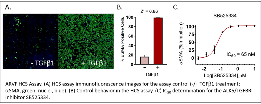
Cell-based fibrosis assays
TGFb1 stimulated myofibroblast differentiation
•Alpha smooth muscle actin (aSMA) primary readout
•Secondary readouts (e.g. collagen): cell-dependent
•Current assays: Adult rat ventricular fibroblasts (ARVF), normal human cardiac fibroblasts (NHCF), normal human lung fibroblast (NHLF)
•Custom assay development possible with your cells
(Ask about our free initial evaluation)
Small molecule collection (~ 3,000)
•FDA & internationally approved drugs
•Target selective (e.g. kinase)
