Anschutz Health Sciences Building. Photo courtesy of ZGF Architects © Connie Zhou
PRECLINICAL IMAGING CONSORTIUM 2024
“Imaging Behind The Barrier, And Beyond — A Life In A BSL Facility”
May 29-31, 2024
CU Anschutz Medical Campus
The Don Elliman Conference Center, located on the 2nd floor of Anschutz Health Science Building (AHSB)
Aurora, CO
Hosted by the University of Colorado Cancer Center Animal Imaging Shared Resources
Organizers: Dr. Natalie Serkova, Jenna Steiner, and Dr. Soudabeh Kargar
Pre-Conference PET/MR Workshop on the morning of May 29th
(more details will be announced soon)
Shared Content Block:
Cancer Center Styles -- Give headings inside accordions and tabs more top margin
Is this your first time attending the Preclinical Imaging Consortium (PIC)? Below is a brief description of the format. Please revisit this page as we continue updating the details of the event. You will see “To Be Announced (TBA)” for the details that will be updated soon.
Conference Location
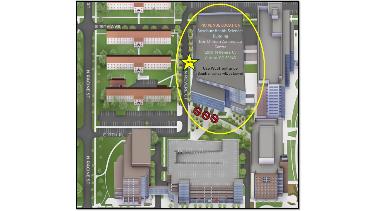
The conference will held be at the University of Colorado Anschutz Medical campus in Aurora, Colorado, in the Don Elliman Conference Center, located on the 2nd floor of Anschutz Health Science Building (AHSB).
Registration and Sponsors
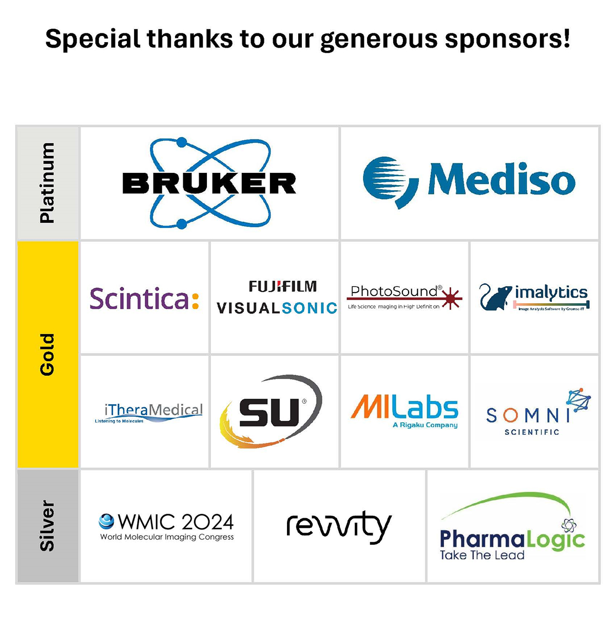
Registration is free for attendees from non-profit organizations, thank you to the generous support of our sponsors. Registration materials will be available from 10:00 AM on Wednesday, May 29, on the 2nd floor of the Anschutz Health Sciences Building (AHSB).
Gala Dinner and Reception
We will hold a gala dinner on site in the Tstreet Kitchen and Cafe.
Vendor “Booths”
Posters and vendor “booths” are in rooms next to the conference and the site of Thursday’s evening reception. Unlike large conferences like WMIC, ISMRM, and RSNA, the vendors do not have enough space for demo equipment. This conference is designed to be smaller and more intimate. All attendees, including vendors, can participate in the talks. Academic attendees are encouraged to talk to the vendors during the breaks. Therefore, the vendors should be near their tables during breaks (though this is not required).
Poster Sessions
There are three types of posters: Industry, Research and Facility.
Industry posters can be strictly marketing or follow a more traditional conference scientific communication approach.
The research/facility posters are intended to communicate a research project that has a significant imaging component that was conducted in an imaging center.
Please send your poster title to: [email protected]. The cork boards are landscape orientation, and the dimensions are 57"W x 0.75"D x 33.5"H. Push pins will be available for you. PhD Posters is offering local printing with on-site delivery. Place your order through this link by 8pm on Monday, May 27th, and it will be delivered directly to the poster session location in plenty of time for setup on Wednesday the 29th. Please contact [email protected] with any questions.
You will be able to vote for your favorite Research and Facility posters.
Facility Tours
This year, you will have the option to tour the Animal Imaging facility, Colorado Animal Imaging SR (BSL2 and BSL3), University of Colorado Research Imaging Center (CU-RIC), PharmaLogic Radiopharmacy and Cyclotron Facility (sign-up).
Exit Meeting (invitation only)
The exit meeting is intended to facilitate the transfer of information from the current host to the next host, and to initiate planning for the next year’s meeting. If you are interested in hosting PIC in the future, contact Jenna Steiner, [email protected] ,for information about the Program Committee Exit Meeting.
Presenter Information
Speakers: Please check the program for the time of your talk. A PC will be available along with a widescreen projector. For Mac users, an HDMI cable with various adapters is available. PowerPoint slides are encouraged. Please give your PowerPoint slides to the AV staff during registration material pick up. It is recommended that you also bring any videos as separate files.
Vendor Presentations
Sponsors will be assigned a table (all), and space for one or more large posters (Gold and Platinum). Gold and Platinum vendors may participate in the Power Pitch session, to be held Thursday, May 30, adjacent to the vendor poster session, where they will present a five-minute overview of their products or announce new equipment. The goal of the power pitch is to make attendees aware of which vendors they may be interested in speaking with in more depth during the conference. Time limits will be strictly enforced!
Air Travel
Aurora can be reached by flying into Denver International Airport (DEN).
Ground Transportation from DEN
Taxi/Uber/Lyft: from the airport to campus or hotel will cost ~ $25-$45.
Rental car: several rental companies at the DEN airport
Hotel
Hyatt Regency Aurora-Denver Conference Center
13200 East 14th Place
Aurora, CO 80011, USA
(720) 925-5314
Last Day to Book: May 10, 2024
Complimentary parking is available for hotel guests.
Conference Location
Anschutz Health Sciences Building
1890 N Revere Ct
Aurora, CO 80045
You can download the program agenda here:
Program Agenda
2024 Preclinical Imaging Consortium Invited Speakers
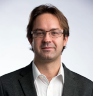 Richard KP Benninger, Ph.D.
Professor of Bioengineering and Pediatrics
Barbara Davis Center for Diabetes
University of Colorado Denver | Anschutz Medical Campus
Dr. Benninger received his PhD in Physics from Imperial College London and then completed postdoctoral training at Vanderbilt University in the laboratory of David Piston in Molecular Physiology and Biophysics. He became an Assistant Professor in the Department of Bioengineering at the University of Colorado Denver | Anschutz medical campus in 2011, establishing his laboratory at the Barbara Davis center for childhood diabetes. His research has focused on applying imaging technologies and biophysical approaches towards understanding how emergent multicellular dynamics govern how insulin and glucagon secretion from the endocrine pancreas coordinates energy metabolism. He research has also focused on developing ultrasound imaging diagnostics for type1 diabetes, the focus for his talk today.
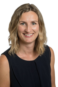 Keara Boss, DVM, Ph.D.
Associate Professor, Flint Animal Cancer Center,
Department of Clinical Sciences, Colorado State University
Dr. Keara Boss attended UMass-Amherst for her undergraduate degree in Animal Science, followed by veterinary school at North Carolina State University. She completed a small animal rotating internship at the University of Pennsylvania and then returned to NC State for residency in radiation oncology. After her residency, she completed a PhD program through NC State, performing the research in radiobiology at Duke University. She then joined the Flint Animal Cancer Center at Colorado State University as a faculty member in the radiation oncology service and she is currently an Associate Professor in the Department of Clinical Sciences.
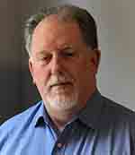 Jim Delikatny, Ph.D.
Research Professor,, Department of Radiology
The University of Pennsylvania
Dr. Delikatny is a Research Professor in the Department of Radiology at the University of Pennsylvania, Director of the Small Animal Imaging Facility and Director of Translational Research for the Center for Precision Surgery . He oversees an interdisciplinary research program on cancer imaging and pharmacology research within the division of Molecular Imaging. His research interests are in noninvasive detection of lipid metabolic and environmental changes associated with tumor progression and response to therapy. He focuses on the development and translation of targeted near-infrared (NIR) and short-wave infrared (SWIR) contrast agents for optical imaging for fluorescence guided surgery.”
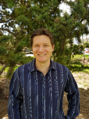 Joe Dragavon, Ph.D.
Director
BioFrontiers Advanced Light Microscopy Core
JSCBB C315
University of Colorado Boulder
Dr. Joe Dragavon has been the Director of the BioFrontiers Institute’s Advanced Light Microscopy Core at the CU Boulder since 2014. Recently, in January 2023, Joe became Director of Core Facilities and Shared Instrumentation within the CU Boulder Research and Innovation Office. Joe is a big supporter of Core Facilities, working within shared resource centers since 2009. He is an active member of the Association for Biomolecular Research Facilities (ABRF) since 2015 and is currently part of their Executive Board.
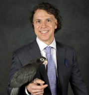 M. Scott Echols, DVM, Diplomate ABVP- Avian Practice
Mobile Avian Surgical Services
The Medical Center for Birds
Dr. Scott Echols is a bird specialist microsurgeon veterinarian living and practicing in Salt Lake City, UT. He is an internationally recognized author, speaker, inventor, visiting professor and Texas A&M College of Veterinary Medicine adjunct professor and distinguished alumnus. His inventions apply to imaging, contrast agents and preservation technologies that are being used to better understand the natural world around us and improve medical diagnostics and treatments. He works in both animal and human medicine in research, teaching, and clinical applications. Several of his artworks have been featured on magazine covers and in art galleries.
 Chad R. Haney, Ph.D.
Center for Advanced Molecular Imaging in the Chemistry Life Processes Institute at Northwestern University
Courtesy appointments in Radiology and Biomedical Engineering
Dr. Haney is a biomedical engineer and has been working on multi-modality imaging for over a decade. His area of research uses medical imaging to identify image-based biomarkers as a means to quantify response to cancer treatment. In his role as managing director of the Center for Advanced Molecular Imaging, he leverages his expertise in medical imaging in other disease models, helping other investigators visualize severity of disease or response to therapy. Each imaging modality has its strengths and weaknesses. Combining more than one imaging modality takes advantage of its strengths while ameliorating its weaknesses. Our goal is to provide quantitative, translational imaging to a diverse group of scientists and clinicians representing numerous disciplines.
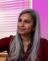 Jayashree Kalpathy-Cramer, Ph.D.
Endowed chair, Ophthalmic data sciences and the founding chief of the Division of Artificial Medical Intelligence,
Department of Ophthalmology at the University of Colorado (CU) School of Medicine
Dr. Jayashree Kalpathy-Cramer is an endowed chair in Ophthalmic data sciences and the founding chief of the Division of Artificial Medical Intelligence in the Department of Ophthalmology at the University of Colorado (CU) School of Medicine. She leads the development and translation of novel artificial intelligence (AI) methods into effective patient care practices at the Sue Anschutz-Rodgers Eye Center. Previously, she was an associate Professor of Radiology at Harvard Medical School where she was actively involved in data science activities with a focus on medical imaging. Her research interests span the spectrum from novel algorithm development to clinical deployment. She is passionate about the potential that machine learning and artificial intelligence have to improve the access and the quality of healthcare in the US and worldwide. Dr. Kalpathy-Cramer has authored over 250 peer-reviewed publications, has written over a dozen book chapters and is a co-inventor on a 15 patents. She graduated from IIT Bombay, India, with a degree in electrical engineering and received her PhD from Rensselaer Polytechnic Institute, also in Electrical Engineering. She returned to academia after almost a decade in the semiconductor industry with a research pivot towards healthcare.
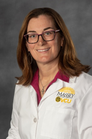 Jennifer Koblinski, Ph.D.
Associate Professor
Virginia Commonwealth University
Director, Cancer Mouse Models Core
CoDirector, Tissue and Data Acquisition and Analysis Core
Department of Pathology
VCU Massey Comprehensive Cancer Center
Dr. Koblinski is an Associate Professor of Pathology at Virginia Commonwealth University and a member of Massey Comprehensive Cancer Center (MCCC), where she serves as the Director of the Cancer Mouse Models Core and Codirector of the Tissue, Data, Acquisition, and Analysis Core. Her research is characterized by a collaborative team approach, focusing on comprehensively understanding cancer growth and progression. She is particularly dedicated to developing targeted therapies for cancer subtypes that lack precision therapy options.
Notably, VCU MCCC recently secured a U54 grant to advance the development of minority patient-derived xenografts (PDX) and organoids (PDO). Dr. Koblinski plays a pivotal role as the co-lead of the PDX/PDO core, contributing her expertise to this initiative.
Her laboratory has made contributions to the field, delving into the role of proteoglycans in cancer. Dr. Koblinski's team utilizes quantitative immunohistochemistry and mouse model imaging in cancer models, showcasing their commitment to rigorous and precise research methods. Additionally, they have experience in developing and validating orthotopic neuroblastoma and breast cancer models.
Dr. Koblinski's work exemplifies her dedication to advancing our understanding of cancer biology and developing innovative therapeutic strategies for cancer subtypes with no precision options.
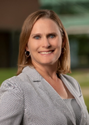 Jori Leszczynski, DVM, DACLAM
Assistant Vice Chancellor for Animal Resources
Director & University Veterinarian
Office of Laboratory Animal Resources
Professor of Pathology, School of Medicine
University of Colorado Denver | Anschutz Medical Campus
Dr. Jori Leszczynski, DVM, DACLAM is currently the Assistant Vice Chancellor for Animal Resources, Director of the Office of Laboratory Animal Resources and University Veterinarian at the University of Colorado Denver | Anschutz Medical Campus, as well as a Professor in the Department of Pathology in the School of Medicine and is a Diplomate of the American College of Laboratory Animal Medicine. Dr. Leszczynski attended The Ohio State University for her Bachelors and Veterinary degrees and performed her residency in Laboratory Animal Medicine at the University of Illinois at Chicago. Throughout her career, she has held multiple leadership roles in animal facilities including Associate Director at the Cleveland Clinic Foundation, Director and AV at National Jewish Health, and Interim Director and AV at the University of Colorado Boulder. In all of her positions, Dr. Leszczynski has focused on program compliance, personnel management, budget, husbandry, and clinical management of the species she oversees. Dr. Leszczynski has a history of service to many professional organizations including leadership roles for the American Society of Laboratory Animal Practitioners (ASLAP, Board of Directors and Membership Committee Chair), American Association for Laboratory Animal Science (AAALAS, 2025-2026 President, Institute for Laboratory Animal Management Committee, Local Arrangements Chair for 2019 Denver AALAS National Meeting, President of the Mile High Branch AALAS), the American College of Laboratory Animal Medicine (ACLAM, Board of Directors) and Public Responsibility in Medicine and Research (PRIM&R, IACUC Conference Planning Committee (Chair), Board of Directors (Chair and Treasurer). She has also served as an ad hoc site visitor for AAALAC International since 2018.
-lo-ph-d.jpg?sfvrsn=cd9cd9bb_1) Su-Tang (Tom) Lo, Ph.D.
Su-Tang (Tom) Lo, Ph.D.
Associate Director in Molecular Imaging
RayzeBio a Bristol Myers Squibb company
With over 18 years of experience in radiochemistry and molecular imaging, I am passionate about applying my skills and knowledge to the development of novel and effective targeted radionuclide therapies for cancer patients. I have a Ph.D. in Radiochemistry and Radiological Science from The University of Texas Southwestern Medical Center at Dallas. Currently, as the Associate Director of Molecular Imaging at RayzeBio, I lead preclinical and translational imaging studies to evaluate the target engagement, tumor profiling, and therapeutic efficacy for multiple projects. My goal is to leverage the power of imaging to enable precision medicine and improve the outcomes for cancer patients.
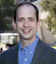 Chris Manuel, DVM, PhD, DACLAM
Senior Associate Director, Office of Laboratory Animal Resources
University of Colorado Denver | Anschutz Medical Campus
Dr. Chris Manuel received his undergraduate degree in Microbiology and Doctorate of Veterinary Medicine from North Carolina State University. Following an internship in emergency veterinary medicine at Florida Veterinary Specialists in Tampa, he performed his residency in Comparative Medicine and obtained his Ph.D. in radiopharmaceutical development at the University of Missouri. He is now the Senior Associate Director of the Office of Laboratory Animal Resources, and Associate Professor in the Department of Pathology at the University of Colorado Denver | Anschutz Medical Campus. He is also the Attending Veterinary for the Rocky Mountain Regional VA Medical Center located in Aurora, CO.
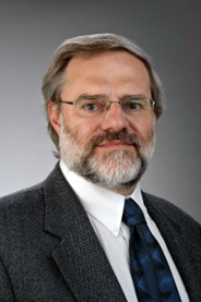 Ralph P. Mason, Ph.D., CSci., CChem., FRSC, FISMRM
Professor of Radiology
University of Texas Southwestern
Director of the Small Animal Imaging Shared Resource (SAISR) at Simmons Comprehensive Cancer Center
Faculty member for graduate programs in Biomedical Engineering and Cancer Biology
I serve as the Director of the Small Animal Imaging Shared Resource (SAISR) at Simmons Comprehensive Cancer Center (SCCC). I am also the Director of the Cancer Imaging Program (CIP) at UT Southwestern Medical Center (UTSW) and a Professor of Radiology. I have over 30 years’ experience in cancer imaging, therapy, and tumor pathophysiology. My primary research interest is prognostic radiology - developing and implementing methods for predicting optimal cancer therapy and assessing early response to treatment ("precision medicine"). My research relates to tumor pathophysiology and developing methods for non-invasive assessment. I am a member of the faculty in graduate programs for Biomedical Engineering, and Cancer Biology. I have advised multiple graduate students, post docs and young faculty, who have gone on to establish productive careers. I collaborate closely with diverse investigators at UT Southwestern and local Institutions (Baylor Univ., Southern Methodist Univ., UT Arlington) in pre-clinical studies and human investigations. I have experience with diverse imaging modalities and have published extensively using MRI and optical imaging as well as ultrasound, PET and most recently photoacoustics. Oxygen-sensitive imaging, hypoxia and assessment of novel therapies are of particular interest to me. Dr. Mason has over 175 peer-reviewed publications with over 12,000 citations and a Google h-index of 61.
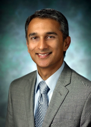 Arvind P. Pathak, Ph.D.
Professor
Depts. of Radiology, Biomedical and Electrical Engineering
The Sidney Kimmel Comprehensive Cancer Center
Institute for NanoBioTechnology
Institute for Computational Medicine
Translational Tissue Engineering Center
The Johns Hopkins University School of Medicine
Dr. Pathak is a global pioneer in the development of hardware, software and “wetware” tools for functional and molecular imaging in health and disease. His team works at the interface of engineering. medicine and design to gain insights into disease, enable biomarker discovery, and impact health and society. Dr. Pathak (www.pathaklab.org) is a biomedical engineer, ideator and mentor whose mission is to “improve lives via the transformative power of imaging”. He received a BS in Electronics Engineering from the University of Poona, India. He received his PhD from the joint program in Functional Imaging between the Medical College of Wisconsin and Marquette University, where he was a Whitaker Foundation Fellow. He completed a postdoctoral fellowship in Molecular Imaging in the Dept. of Radiology at the Johns Hopkins University School of Medicine. At Johns Hopkins University (JHU), Dr. Pathak is Professor in the Schools of Medicine (Radiology and Oncology) and Engineering (Biomedical and Electrical Engineering). He is also a member of the Sidney Kimmel Comprehensive Cancer Center, the Institute for NanoBioTechnology (INBT), the Institute for Computational Medicine (ICM) and the Translational Tissue Engineering Center (TTEC). His research in imaging has been recognized by multiple awards such as the Bill Negendank Award from the International Society for Magnetic Resonance in Medicine (ISMRM) given to “outstanding young investigators in cancer MRI” and the Career Catalyst Award from the Susan Komen Foundation. Dr. Pathak has served in multiple leadership roles spearheading imaging initiatives, mentoring junior faculty, review panels for national and international funding agencies, and journal editorial boards. He was the Chair of the Cancer Section for the International Society for Magnetic Resonance in Medicine, and currently serves on the Executive Council of the Microcirculatory Society (MCS) and was the Chair of the NIH’s Emerging Imaging Technologies in Neuroscience (EITN) study section. He directs an interdisciplinary, nurturing and continuously funded laboratory, and has mentored over 100 award-winning (e.g. Fulbright and Howard Hughes Fellows) students. He is dedicated to mentoring the next generation of thought leaders and was the recipient of the ISMRM’s Outstanding Teacher Award in 2014, was a JHU Career Champion nominee in 2018, and recognized as a 125 Hopkins Hero by the JHU School of Medicine for his “outstanding dedication to the core values of Johns Hopkins University”.
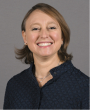 Federica Pisaneschi, Ph.D.
Assistant Professor, Center for Translational Cancer Research at the Brown Foundation Institute of Molecular Medicine for the Prevention of Human Diseases (IMM),
The University of Texas Health Science Center at Houston
Dr. Federica Pisaneschi is currently an Assistant Professor in the Center for Translational Cancer Research at the Brown Foundation Institute of Molecular Medicine for the Prevention of Human Diseases (IMM) at The University of Texas Health Science Center at Houston.
Dr. Pisaneschi started her career as a synthetic organic chemist earning her master’s degree and Ph.D. at the University of Florence (Italy). She then remained in Florence with a postdoctoral fellowship before being hired as a postdoctoral research associate at Hammersmith Hospital, Imperial College London (UK). At Imperial College, she worked at the Comprehensive Cancer Imaging Center (CCIC), where she developed several PET agents. Among those, 18F-Fluoropivalic acid, a report of lipid metabolism, has advanced to the clinic and is currently in Phase IIa-III clinical trial. In 2015, she moved to the USA and joined MD Anderson Cancer Center where she worked until 2023. At MD Anderson, she continued in the research and development of new radiopharmaceuticals, enabled R&D radiochemistry research, and helped faculty add radiochemistry and PET to their programs. At MD Anderson, 4-[18F] fluoronaphthol, a first in class reporter on myeloid cell activation and the topics of today’s talk, was developed. 18F-4FN is currently in Phase 0-I clinical trial.
Her research is focused on expanding the use of PET to guide personalized medicine by developing new radiopharmaceuticals and using known, less-common radiopharmaceuticals in mechanism-based innovative molecular imaging applications. She has successfully contributed to the field of chemistry, radiochemistry and radiopharmaceuticals discovery with 50+ peer-reviewed publications, seven patents and 80+ reports at international meetings. She is deeply involved with the World Molecular Imaging Society (WMIS) and the Society of Radiopharmaceutical Sciences (SRS), especially regarding education.
She is now building a new team of radiopharmaceuticals developers and molecular imagers at UTHealth Houston. Current projects in her lab are focused on imaging of neuroinflammation and neurodegeneration, phenotyping metabolic reprograming of prostate cancer, targeted radiotherapy and development of novel 18F-labeling strategies.
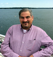 Charles A. Scanga, Ph.D.
University of Pittsburgh School of Medicine, Department of Microbiology and Molecular Genetics.
University of Pittsburgh’s Center for Vaccine Research.
Dr. Scanga received his Ph.D. in Microbiology and Molecular Virology in 2000 from the University of Pittsburgh, where he studied latent TB in mouse models in the lab of JoAnne Flynn. He did a post-doctoral fellowship in Alan Sher’s lab at NIH, where he studied toxoplasma, TB, and HIV- M. tuberculosis co-infections. In 2005, he joined the Aeras Global TB Vaccine Foundation and led the Animal Studies section which evaluated novel TB vaccine candidates in a variety of animal models. He returned to Pitt in 2008 and is currently a Research Associate Professor. His primary research interest is HIV - M. tuberculosis co-infection. He also works closely with Dr. JoAnne Flynn, serving as the project manager for her TB studies on diverse topics including TB vaccines, anti-TB drugs, PET probes for infectious diseases, and basic TB pathogenesis. As the manager of the PET/CT imaging facility within the Regional Biocontainment Laboratory in Pitt’s Center for Vaccine Research, Dr. Scanga works closely with other Pitt researchers to apply PET/CT to research on various other microbial pathogens.
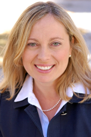 Natalie J. Serkova, MSc, Ph.D.
Charles L. Adams Professor and Vice Chair for Research
Department of Radiology
Associate Director (Shared Resources), CU Cancer Center
Director, Colorado Animal Imaging Shared Resource (AISR)
Director, CCTSI Pilot Grant Program
Dr. Serkova is a tenured Professor and Vice Chair Research of Radiology at the University of Colorado Anschutz Medical Campus. She is the founding director of Colorado Animal Imaging Shared Resource, which is supported by the NIH/ NCI, the Cancer Center and the Colorado Clinical Translational Sciences Institute (CCTSI). Natalie’s formal education includes training in Physics at the University of Grenoble (France, BS) and Biophysics/ Biochemistry the University of Kiev (Ukraine, MSc). After completing her Ph.D. in MR Physics, MRI Division, University of Bremen (Germany), she joined the Departments of Biopharmaceutical Sciences at the University of California at San Francisco (UCSF) and Radiology at Stanford University as a postdoctoral fellow in 1997. There, she was trained in animal imaging PK/PD. Dr. Serkova’s research interests are in the development of new imaging probes and acquisition protocols for animal cancer models using the state-of-the-art preclinical scanners (MRI, CT, PET/ SPECT, optical imaging). She has mentored 37 undergraduate students, 15 graduate students, 7 post-doctoral students and residents and published over 160 peer-reviewed publications in the area of oncologic imaging and metabolism.
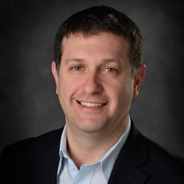 Erik M. Shapiro, MS, Ph.D.
Associate Chair of Research, Department of Radiology
Professor of Radiology, Physiology, Chemical Engineering and Material Science, and Biomedical Engineering
Division Chief, Biomedical Imaging
Institute for Quantitative Health Sciences and Engineering
Erik Shapiro is Associate Chair of Research and a professor in the Department of Radiology. His Molecular and Cellular Imaging Laboratory (MCIL) develops and uses magnetic resonance imaging (MRI), positron emission tomography (PET) and x-ray computed tomography (CT) for molecular and cellular imaging of biological phenomena, regenerative medicine, and early detection of disease. Dr. Shapiro graduated from the State University of New York at Binghamton with a degree in chemistry and holds a MS and Ph.D. in chemistry from the University of Pennsylvania. He conducted his post-doctoral work in molecular imaging at the National Institutes of Health. He is a recipient of an NIH Director's New Innovator Award as well as a Distinguished Investigator Award from the Radiological Society of North America.
 Rajesh Uthamanthil, Ph.D.
Professor (Affiliate), Department of Comparative Medicine Chief Medical Officer (Preclinical/Translational)
Seattle Children’s Research Institute University of Washington
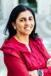 Pavithra Viswanath, Ph.D.
Associate Professor Department of Radiology and Biomedical Imaging,
University of California San Francisco
Dr. Pavithra Viswanath is an Associate Professor in the Department of Radiology and Biomedical Imaging at the University of California San Francisco. The overarching goal of her research is to leverage tumor metabolism for the identification of novel, clinically translatable metabolic imaging biomarkers and therapeutic vulnerabilities for brain tumors. To this end, her research program utilizes advanced MRS/I methods, including 1H-, hyperpolarized 13C-, and deuterium MRS in cell and animal models for imaging oncogenic events in brain tumors. Dr. Viswanath received a Ph.D. in Biochemistry at the Indian Institute of Science, Bangalore, India. She is the recipient of numerous NIH R01, R21, P50 program project, Department of Defense, and philanthropic grants focused on brain tumor imaging and therapy.
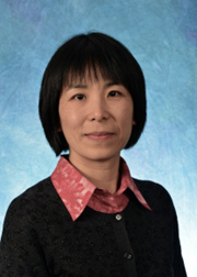 Hong Yuan, Ph.D.
Department of Radiology,
University Of North Carolina at Chapel Hill
Dr. Hong Yuan is a Research Professor in the Department of Radiology and the Director of Small Animal Imaging Facility at University of North Carolina Chapel Hill. Dr. Yuan is trained in biomedical engineering, and has extensive experience in multimodality imaging in preclinical setting. Her areas of interest include molecular imaging of tumor microenvironment, image-guided radiotherapy, and radiation induced normal tissue injury.
You can view programs from previous PIC meetings from the list below:
2023 PIC Hosted by Michigan State University, East Lansing, MI, 2023
2019 MPIC Hosted by Northwestern University, Evanston, IL 2019
MPIC Hosted by Washington University, St. Louis, MO 2018
MPIC Hosted by Vanderbilt University, Nashville, TN 2017
MPIC Hosted by University of Wisconsin-Madison, Madison, WI 2016