Heart Valve Disorders
What Do Heart Valves Do?
Heart valves help regulate the flow of blood from your body, into the lungs and back out to your body. The four chambers of the heart are separated by the use of valves that orchestrate which direction blood is sent and coordinate the timing of your circulatory system. It is imperative that these valves open and close at the appropriate times or the resulting dysfunction can lead to a lack of oxygen being sent around your body. Each valve has two responsibilities: to open correctly and to close correctly.
Heart Chambers and Valves
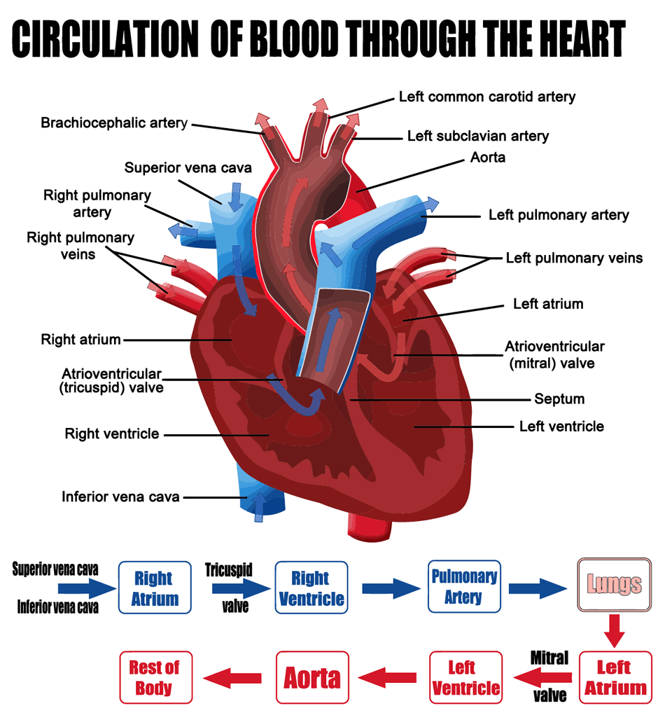
It is helpful to understand the parts of the heart and the role of each valve when learning about heart valve disease. The heart has two sides and four chambers, two on each side that hold the blood while it is pumped through the heart. The two upper chambers are the right and left atria, and the lower chambers are the right and left ventricles. (See diagram below.) Blood enters the heart into the right atrium, through the tricuspid valve and into the right ventricle. From here is passes through the pulmonary valve and into the lungs to receive oxygen. Upon return from the lungs, blood flows into the left atrium through the mitral valve and into the left ventricle. Finally the blood is pumped through the aortic valve and back into the body.
It's Not Always a Perfect System
Sometimes there are problems with the flow of blood through your heart. There is a variety of conditions and diseases that can lead to complications with heart chambers and valves. Additionally there are many different ways to treat or repair valve problems.

It goes without saying that problems with the function of your heart valves can be serious and even fatal. There are several types of common valve problems and many different causes for problems associated with their efficiency.
Valve Stenosis
Stenosis is the term used to describe a valve that does not open correctly. At times, the valves flaps (also called leaflets) can thicken or even fuse together impeding their ability to open and close as necessary to allow for effective blood flow. This can lead to a lack of oxygenated blood flowing into the body. Stenosis can be caused by age. Rarely, people can be born with a malformed valve that doesn't open and close properly.
Aortic valve stenosis is one of the most common and serious valve problems. Most commonly, aortic valve stenosis develops in the elderly through the buildup of calcium around the valve. Some people are born with aortic stenosis as a congenital heart defect.
Pulmonary valve stenosis is most commonly a result of a congenital heart defect. Because of this, it is rare in adults as it is most commonly diagnosed in childhood by the presence of a murmur.
Mitral valve and tricuspid valve stenosis are less common and almost always result from rheumatic fever, occurring after a bought of untreated streptococcal infection i.e. "strep throat". Due to easy access to antibiotics in the United States, these valve diseases are less common.
Mitral Valve Prolapse
Mitral valve prolapse, or MVP, is the term that refers to incorrect function of the two valve flaps in the mitral valve that controls blood flow between the left atrium and ventricle. MVP is frequently without symptoms but can be detected as a murmur during a routine physical exam. In most cases, it's harmless. According to the American Heart Association, MVP occurs in 2 to 3 percent of the population.
Mitral valve syndrome is the term coined for those that do experience symptoms of MVP. Those symptoms include chest pain, dizziness, fatigue, heart palpitations or shortness of breath. When leakage though the mitral valve becomes severe, it may require mitral valve surgery for repair or replacement.
Heart Valve Regurgitation
Valve regurgitation is also known as leaking heart valves. This occurs when the valve leaflets are closing and blood flows back trough the valve. It can also happen when the valves are entirely closed but they allow flood to leak. A leaking valve forces the heart to work harder to pump blood. Depending on the severity of the valve leak, it may need to be repaired or replaced surgically.
Symptoms
It is crucial that your doctor listen to your heart to check for possible symptoms resulting from undiagnosed heart valve disease. Some people do not have or ever notice any symptoms and their condition is only discovered through the detection of a murmur during a routine physical examination. Some symptoms you may experience are:
- Chest pain
- Palpitations
- Fatigue
- Lightheadedness
- Shortness of breath
- Syncope
Risks
Heart valve disease is most common in older people and is becoming more prevalent as life expectancy increases due to improved medical care and knowledge of disease treatment. Heart valves become lined with calcium deposits and the leaflets thicken and become stiffer over time. Risk for valve disease is increased in those that have had rheumatic fever or infective endocarditis. Additionally, a history of heart attack, heart failure, arrhythmia or congenital heart defects can increase your risk for developing valve disease.
Echocardiography
Echocardiography is the main tool used to evaluate heart valve disease. This procedure, sometimes referred to as an echo test or heart ultrasound, takes real time moving pictures of your heart as it works. These video images can be evaluated by your doctor to view heart valvefunction. This procedure does not require a hospital stay and is painless.
Cardiac Catheterization
Cardiac catheterization is performed to confirm the presence of a suspected heart ailment and can be a diagnostic tool or used interventionally to treat a heart condition. A cardiac catheterization is a broad term used to describe the insertion of a catheter into an artery to gain access to the heart; this is generally done through the femoral or radial arteries under a local anesthetic. Contrast agents can; be used to observe blood flow and heart valve function.
Chest X-ray
A chest x-ray can be used to view much of the anatomic structure of the chest including the lungs and heart. This procedure is very common and is painless.
Electrocardiogram
An electrocardiogram, also called an EKG or ECG, is used to assess the electrical activity of the heart over a period of time. This is done by placing electrodes, or leads, on the surface of the body that can detect abnormalities in heart function including its rhythm and electrical impulses.
The image to the right show how EKG leads are placed on the body. This process is painless and takes just a few minutes.
Exercise Stress Test
An exercise stress test, sometimes called a stress test can be performed in conjunction with echocardiography and an EKG to note changes in your heart's function during exercise. To perform this test, patients are asked to walk on a treadmill to increase heart rate during and after which its activity can be observed through ultrasound and EKG.
If it is determined that you have an abnormality with one or several of your heart valves, you and your physician can discuss potential treatment options or surgery.
The presence of problems in heart valve function is usually diagnosed first through auscultation, or listening to the heart with a stethoscope. The detection of a possible abnormality will incent your physician to request additional testing.
Heart Valve Repair/Replacement
Inside the heart are four valves (Pulmonary, Tricuspid, Aortic, and Mitral). When healthy and working properly, these valves keep the blood flowing in the right direction.
Heart valves can lose function due to a variety of causes. Heart valves may not open completely (a condition referred to as stenosis) or they may not close completely (referred to as regurgitation). Heart Valve Disease is associated with a variety of symptoms, and surgical intervention is often the preferred course of treatment.
Valve Repair Surgery
The mitral valve is the most commonly repaired heart valve, but all four of the valves may be repaired (rather than replaced) depending upon the nature and severity of heart-valve disease present.
The most common heart-valve repair techniques are commissurotomy, in which fused valve leaflets (flaps) are separated to widen the opening; decalcification, in which calcium deposits are surgically removed to allow the valve to function properly; and leaflet reshaping, in which poorly functioning sections of a valve leaflet are removed.
Valve Replacement Surgery
Depending upon the nature and severity of heart valve disease, your physician may recommend valve replacement surgery as the most appropriate form of treatment. Disease of the aortic valve and the pulmonic valve are more often treated with valve replacement rather than repair. Valve replacement can involve the use of either mechanical, biological, or allograft valves.
Open Procedures vs. TAVR
Historically, valve replacement surgery has required the use of a sternotomy or the opening of your sternum to gain access to your heart. Currently, many patients are able to receive live saving valve replacement surgery through a minimally invasive procedure called Transcatheter Aortic Valve Replacement or TAVR. This procedure uses only very small incisions as the valve is placed into the heart through a catheter system as opposed to direct visualization. The video below shows an artist rendering of the TAVR procedure being performed.
TAVR
Transcatheter Aortic Valve Replacement (TAVR) is a commonly performed, minimally invasive heart-valve replacement surgery. In TAVR, a new aortic valve is delivered to the site of implantation by way of a catheter. The catheter is typically inserted into the body at the femoral artery in the groin area and guided along this artery to the heart. As an alternative to open heart surgery, this type of heart-valve surgery usually results in a shorter recovery time. It can be particularly beneficial for patients who are too ill to undergo open heart valve-replacement surgery.
The heart is a continuously beating pump that keeps the body supplied with oxygenated blood. The left side of the heart receives oxygenated blood from the lungs and pumps this oxygenated blood out to the body. The right side of the heart receives de-oxygenated blood from the body and pumps it to the lungs where it will be re-oxygenated.
This information is provided by the Department of Surgery at the University of Colorado School of Medicine. It is not intended to replace the medical advice of your doctor or healthcare provider. Please consult your healthcare provider for advice about a specific medical condition.